Pilch Matthäus, Wenner Yaroslava, Strohmayr Elisabeth, Preising Markus, Friedburg Christoph, Meyer Zu Bexten Erdmuthe, Lorenz Birgit, Stieger Knut
Biomed Opt Express. 2012 Jul 1;3(7):1478-91. doi: 10.1364/BOE.3.001478. Epub 2012 Jun 4.
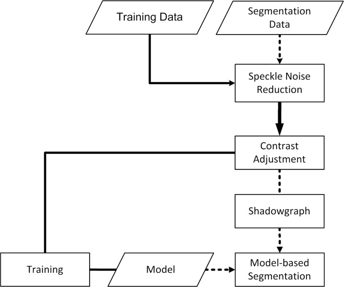
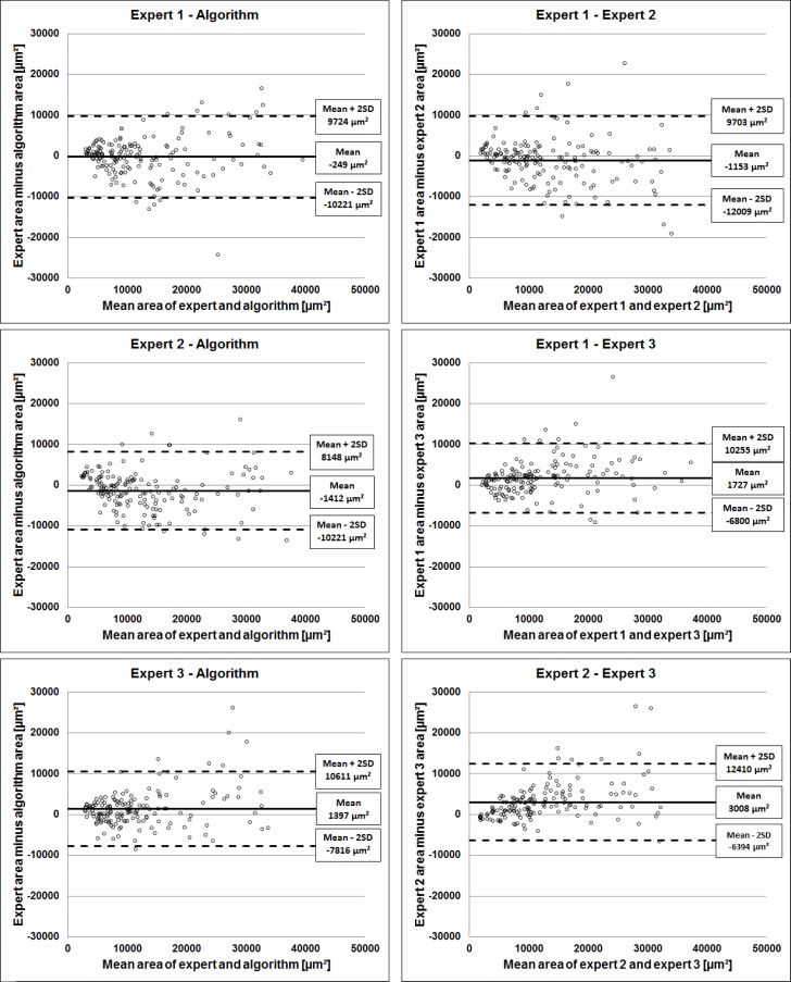
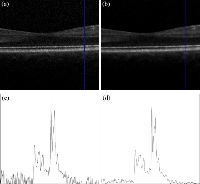
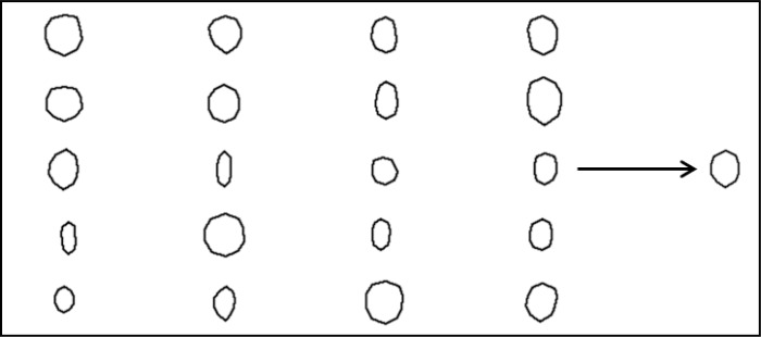
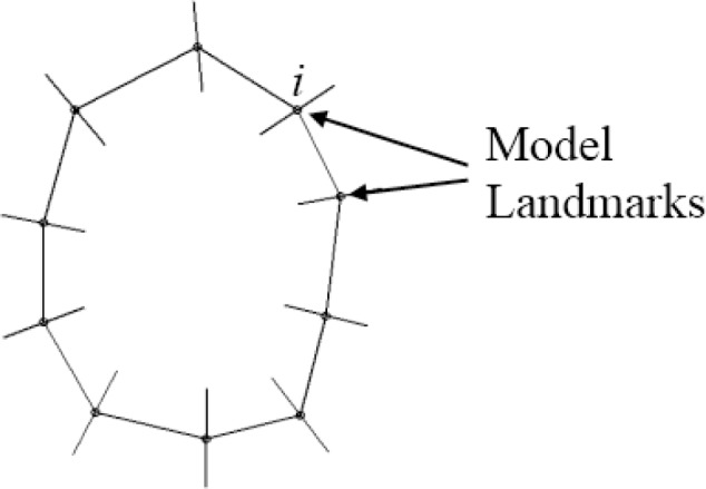
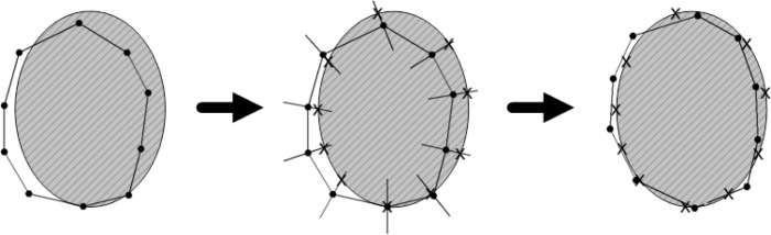
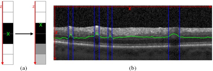
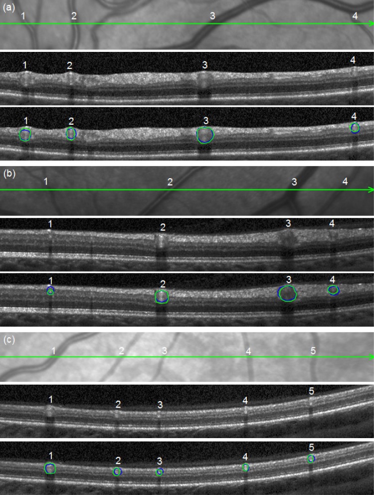
The correct segmentation of blood vessels in optical coherence tomography (OCT) images may be an important requirement for the analysis of intra-retinal layer thickness in human retinal diseases. We developed a shape model based procedure for the automatic segmentation of retinal blood vessels in spectral domain (SD)-OCT scans acquired with the Spectralis OCT system. The segmentation procedure is based on a statistical shape model that has been created through manual segmentation of vessels in a training phase. The actual segmentation procedure is performed after the approximate vessel position has been defined by a shadowgraph that assigns the lateral vessel positions. The active shape model method is subsequently used to segment blood vessel contours in axial direction. The automated segmentation results were validated against the manual segmentation of the same vessels by three expert readers. Manual and automated segmentations of 168 blood vessels from 34 B-scans were analyzed with respect to the deviations in the mean Euclidean distance and surface area. The mean Euclidean distance between the automatically and manually segmented contours (on average 4.0 pixels respectively 20 µm against all three experts) was within the range of the manually marked contours among the three readers (approximately 3.8 pixels respectively 18 µm for all experts). The area deviations between the automated and manual segmentation also lie within the range of the area deviations among the 3 clinical experts. Intra reader variability for the experts was between 0.9 and 0.94. We conclude that the automated segmentation approach is able to segment blood vessels with comparable accuracy as expert readers and will provide a useful tool in vessel analysis of whole C-scans, and in particular in multicenter trials.
在光学相干断层扫描(OCT)图像中正确分割血管,可能是分析人类视网膜疾病中视网膜内层厚度的一项重要要求。我们开发了一种基于形状模型的程序,用于自动分割使用Spectralis OCT系统采集的光谱域(SD)-OCT扫描中的视网膜血管。该分割程序基于一个统计形状模型,该模型是在训练阶段通过手动分割血管创建的。实际分割程序是在通过阴影图确定了大致血管位置(该阴影图确定了血管的横向位置)之后执行的。随后使用主动形状模型方法在轴向分割血管轮廓。三位专家读者将自动分割结果与相同血管的手动分割结果进行了对比验证。针对34次B扫描中的168条血管的手动和自动分割,分析了平均欧几里得距离和表面积的偏差。自动分割轮廓与手动分割轮廓之间的平均欧几里得距离(平均分别为4.0像素,相对于所有三位专家为20µm)在三位读者手动标记轮廓的范围内(所有专家约为3.8像素,分别为18µm)。自动分割与手动分割之间的面积偏差也在三位临床专家的面积偏差范围内。专家读者的读者内变异性在0.9至0.94之间。我们得出结论,自动分割方法能够以与专家读者相当的准确性分割血管,并将为整个C扫描的血管分析,特别是在多中心试验中提供一个有用的工具。