Department of Biology, Dalhousie University, 1355, Oxford Street, Halifax, Nova Scotia B3H 4R2, Canada.
BMC Plant Biol. 2012 Jul 25;12:115. doi: 10.1186/1471-2229-12-115.
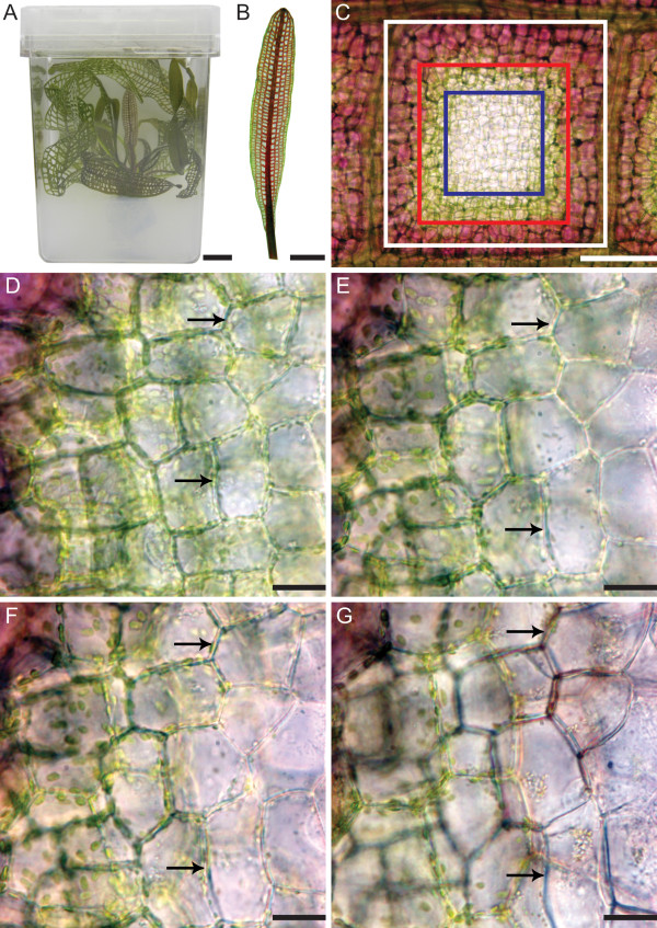
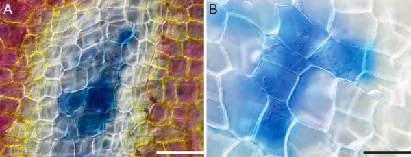
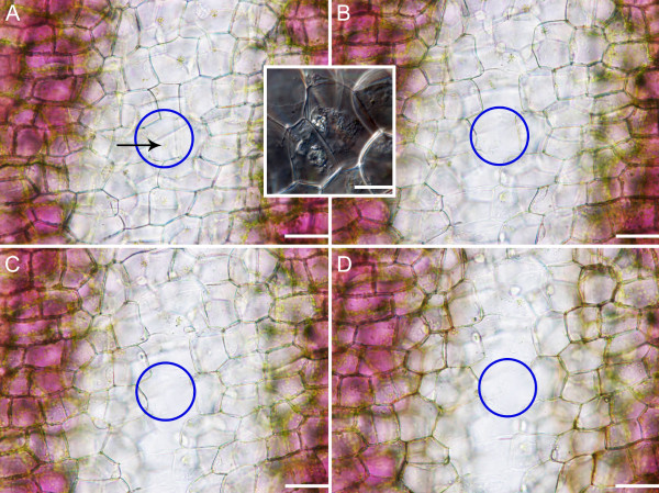
Developmentally regulated programmed cell death (PCD) is the controlled death of cells that occurs throughout the life cycle of both plants and animals. The lace plant (Aponogeton madagascariensis) forms perforations between longitudinal and transverse veins in spaces known as areoles, via developmental PCD; cell death begins in the center of these areoles and develops towards the margin, creating a gradient of PCD. This gradient was examined using both long- and short-term live cell imaging, in addition to histochemical staining, in order to establish the order of cellular events that occur during PCD.
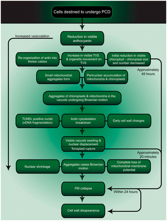
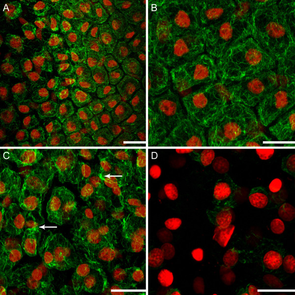
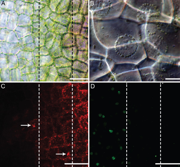
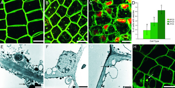
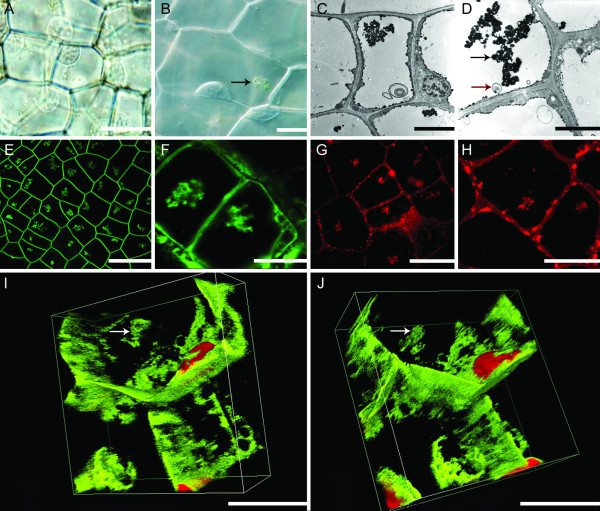
The first visible change observed was the reduction in anthocyanin pigmentation, followed by initial chloroplast changes and the bundling of actin microfilaments. At this stage, an increased number of transvacuolar strands (TVS) was evident. Perhaps concurrently with this, increased numbers of vesicles, small mitochondrial aggregates, and perinuclear accumulation of both chloroplasts and mitochondria were observed. The invagination of the tonoplast membrane and the presence of vesicles, both containing organelle materials, suggested evidence for both micro- and macro-autophagy, respectively. Mitochondrial aggregates, as well as individual chloroplasts were subsequently seen undergoing Brownian motion in the vacuole. Following these changes, fragmentation of nuclear DNA, breakdown of actin microfilaments and early cell wall changes were detected. The vacuole then swelled, causing nuclear displacement towards the plasma membrane (PM) and tonoplast rupture followed closely, indicating mega-autophagy. Subsequent to tonoplast rupture, cessation of Brownian motion occurred, as well as the loss of mitochondrial membrane potential (ΔΨm), nuclear shrinkage and PM collapse. Timing from tonoplast rupture to PM collapse was approximately 20 minutes. The entire process from initial chlorophyll reduction to PM collapse took approximately 48 hours. Approximately six hours following PM collapse, cell wall disappearance began and was nearly complete within 24 hours.
Results showed that a consistent sequence of events occurred during the remodelling of lace plant leaves, which provides an excellent system to study developmental PCD in vivo. These findings can be used to compare and contrast with other developmental PCD examples in plants.
发育调控程序化细胞死亡(PCD)是植物和动物整个生命周期中细胞的可控死亡。马岛水蕹(Aponogeton madagascariensis)通过发育性 PCD 在纵脉和横脉之间的空间形成穿孔,称为气腔;细胞死亡首先发生在这些气腔的中心,然后向边缘发展,形成 PCD 的梯度。通过长期和短期活细胞成像以及组织化学染色来检查这个梯度,以确定 PCD 过程中发生的细胞事件的顺序。
观察到的第一个可见变化是花青素色素沉着的减少,随后是初始叶绿体变化和肌动蛋白微丝的束集。在这个阶段,明显增加了跨液泡链(TVS)的数量。也许与此同时,观察到囊泡、小线粒体聚集体以及叶绿体和线粒体的核周积累的数量增加。液泡膜的内陷和含有细胞器物质的囊泡的存在分别表明存在微自噬和巨自噬的证据。随后,线粒体聚集体以及单个叶绿体在液泡中被观察到布朗运动。在这些变化之后,检测到核 DNA 的片段化、肌动蛋白微丝的断裂和早期细胞壁的变化。然后,液泡肿胀,导致核向质膜(PM)移位,紧随其后的是液泡破裂,表明巨自噬。液泡破裂后,布朗运动停止,线粒体膜电位(ΔΨm)丧失,核收缩和 PM 崩溃。从液泡破裂到 PM 崩溃的时间大约为 20 分钟。从初始叶绿素减少到 PM 崩溃的整个过程大约需要 48 小时。PM 崩溃后大约 6 小时,细胞壁开始消失,24 小时内几乎完全消失。
结果表明,在马岛水蕹叶片的重塑过程中发生了一致的事件序列,为活体研究发育性 PCD 提供了一个极好的系统。这些发现可用于与植物中其他发育性 PCD 实例进行比较和对比。