Department of Hematology-Oncology, Kaohsiung Chang Gung Memorial Hospital and Chang Gung University College of Medicine, Kaohsiung, Taiwan.
BMC Cancer. 2012 Aug 1;12:328. doi: 10.1186/1471-2407-12-328.
Correct detection of bone metastases in patients with esophageal squamous cell carcinoma is pivotal for prognosis and selection of an appropriate treatment regimen. Whole-body bone scan for staging is not routinely recommended in patients with esophageal squamous cell carcinoma. The aim of this study was to investigate the role of bone scan in detecting bone metastases in patients with esophageal squamous cell carcinoma.
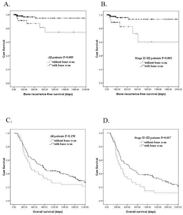
We retrospectively evaluated the radiographic and scintigraphic images of 360 esophageal squamous cell carcinoma patients between 1999 and 2008. Of these 360 patients, 288 patients received bone scan during pretreatment staging, and sensitivity, specificity, positive predictive value, and negative predictive value of bone scan were determined. Of these 360 patients, surgery was performed in 161 patients including 119 patients with preoperative bone scan and 42 patients without preoperative bone scan. Among these 161 patients receiving surgery, 133 patients had stages II + III disease, including 99 patients with preoperative bone scan and 34 patients without preoperative bone scan. Bone recurrence-free survival and overall survival were compared in all 161 patients and 133 stages II + III patients, respectively.
The diagnostic performance for bone metastasis was as follows: sensitivity, 80%; specificity, 90.1%; positive predictive value, 43.5%; and negative predictive value, 97.9%. In all 161 patients receiving surgery, absence of preoperative bone scan was significantly associated with inferior bone recurrence-free survival (P = 0.009, univariately). In multivariate comparison, absence of preoperative bone scan (P = 0.012, odds ratio: 5.053) represented the independent adverse prognosticator for bone recurrence-free survival. In 133 stages II + III patients receiving surgery, absence of preoperative bone scan was significantly associated with inferior bone recurrence-free survival (P = 0.003, univariately) and overall survival (P = 0.037, univariately). In multivariate comparison, absence of preoperative bone scan was independently associated with inferior bone recurrence-free survival (P = 0.009, odds ratio: 5.832) and overall survival (P = 0.029, odds ratio: 1.603).
Absence of preoperative bone scan was significantly associated with inferior bone recurrence-free survival, suggesting that whole-body bone scan should be performed before esophagectomy in patients with esophageal squamous cell carcinoma, especially in patients with advanced stages.
正确检测食管鳞癌患者的骨转移对预后和治疗方案的选择至关重要。全身骨扫描在食管鳞癌患者中并不常规推荐用于分期。本研究旨在探讨骨扫描在检测食管鳞癌患者骨转移中的作用。
我们回顾性评估了 1999 年至 2008 年间 360 例食管鳞癌患者的影像学和闪烁扫描图像。在这 360 例患者中,288 例在治疗前分期时接受了骨扫描,确定了骨扫描的敏感性、特异性、阳性预测值和阴性预测值。在这 360 例患者中,161 例行手术治疗,其中 119 例行术前骨扫描,42 例未行术前骨扫描。在这 161 例接受手术的患者中,133 例为 II+III 期疾病,其中 99 例术前骨扫描,34 例无术前骨扫描。在所有 161 例患者和 133 例 II+III 期患者中,分别比较了骨无复发生存率和总生存率。
骨转移的诊断性能如下:敏感性 80%;特异性 90.1%;阳性预测值 43.5%;阴性预测值 97.9%。在所有接受手术的 161 例患者中,术前无骨扫描与较差的骨无复发生存率显著相关(P=0.009,单变量)。多变量比较时,术前无骨扫描(P=0.012,优势比:5.053)是骨无复发生存的独立不良预后因素。在接受手术的 133 例 II+III 期患者中,术前无骨扫描与较差的骨无复发生存率(P=0.003,单变量)和总生存率(P=0.037,单变量)显著相关。多变量比较时,术前无骨扫描与骨无复发生存率(P=0.009,优势比:5.832)和总生存率(P=0.029,优势比:1.603)显著相关。
术前无骨扫描与较差的骨无复发生存率显著相关,提示全身骨扫描应在食管鳞癌患者行食管切除术前行,特别是在晚期患者中。