San Raffaele Institute Sulmona, L'Aquila, Italy.
PLoS One. 2012;7(7):e42339. doi: 10.1371/journal.pone.0042339. Epub 2012 Jul 31.
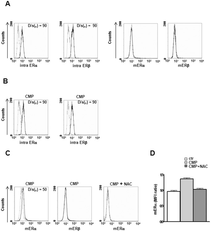
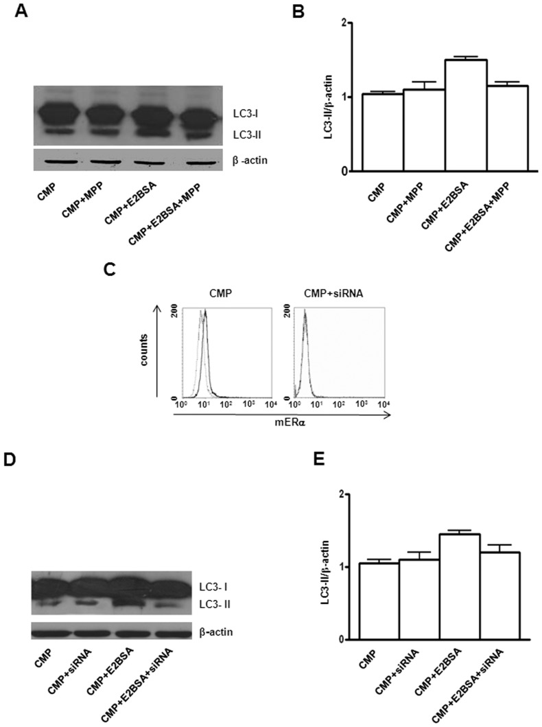
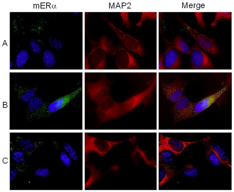
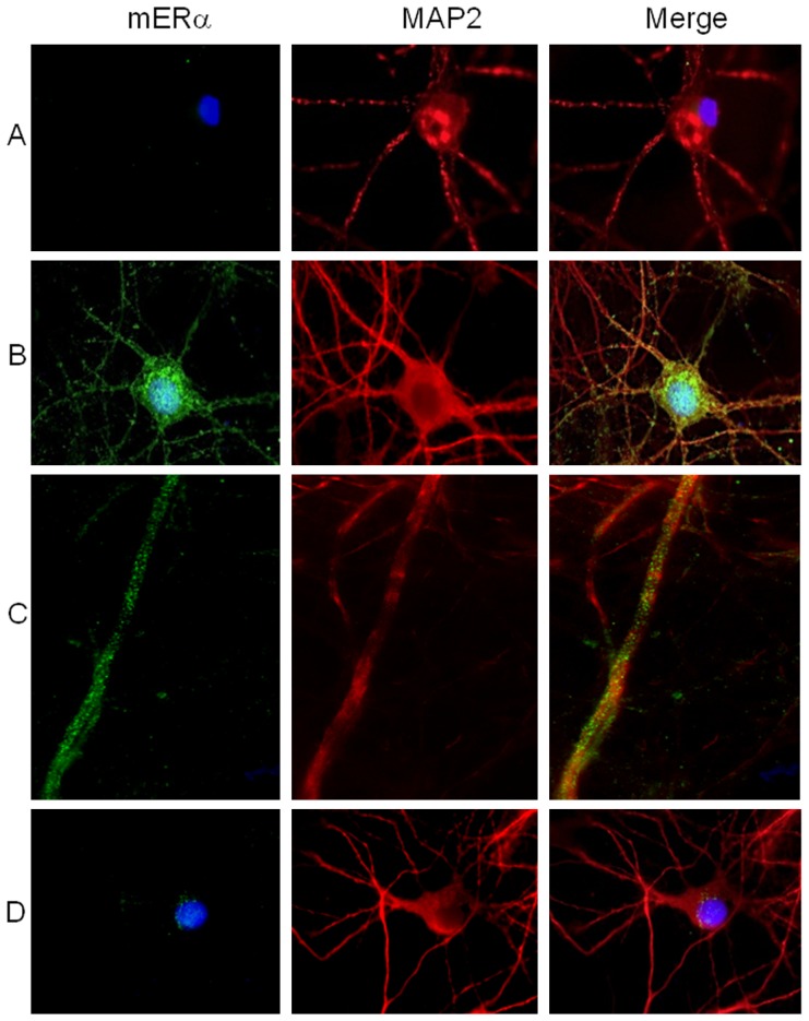
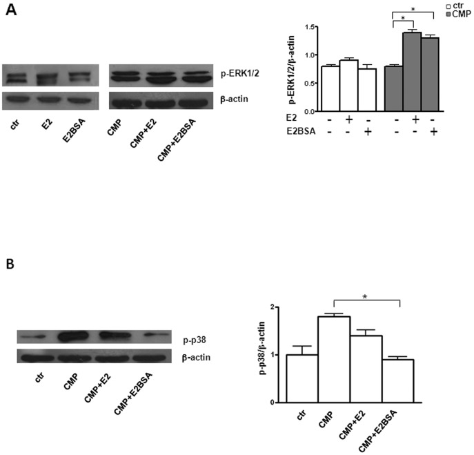
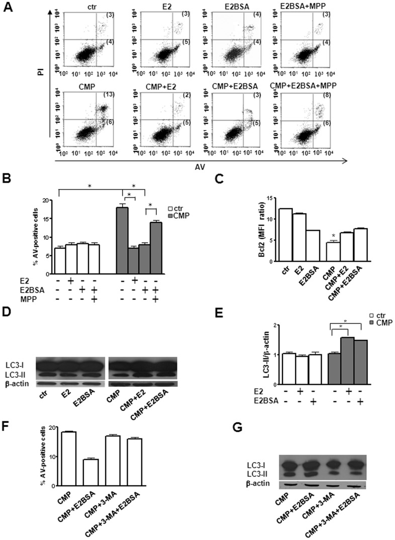
In addition to the classical nuclear estrogen receptor, the expression of non-nuclear estrogen receptors localized to the cell surface membrane (mER) has recently been demonstrated. Estrogen and its receptors have been implicated in the development or progression of numerous neurodegenerative disorders. Furthermore, the pathogenesis of these diseases has been associated with disturbances of two key cellular programs: apoptosis and autophagy. An excess of apoptosis or a defect in autophagy has been implicated in neurodegeneration. The aim of this study was to clarify the role of ER in determining neuronal cell fate and the possible implication of these receptors in regulating either apoptosis or autophagy. The human neuronal cell line SH-SY5Y and mouse neuronal cells in primary culture were thus exposed to chronic minimal peroxide treatment (CMP), a form of subcytotoxic minimal chronic stress previously that mimics multiple aspects of long-term cell stress and represents a limited molecular proxy for neurodegenerative processes. We actually found that either E2 or E2-bovine serum albumin construct (E2BSA, i.e. a non-permeant form of E2) was capable of modulating intracellular cell signals and regulating cell survival and death. In particular, under CMP, the up-regulation of mERα, but not mERβ, was associated with functional signals (ERK phosphorylation and p38 dephosphorylation) compatible with autophagic cytoprotection triggering and leading to cell survival. The mERα trafficking appeared to be independent of the microfilament system cytoskeletal network but was seemingly associated with microtubular apparatus network, i.e., to MAP2 molecular chaperone. Importantly, antioxidant treatments, administration of siRNA to ERα, or the presence of antagonist of ERα hindered these events. These results support that the surface expression of mERα plays a pivotal role in determining cell fate, and that ligand-induced activation of mER signalling exerts a powerful cell-survival signal. These results shed new light on the pathogenetic mechanisms leading to neuronal cell degeneration.
除了经典的核雌激素受体外,最近还证明了定位于细胞膜表面的非核雌激素受体(mER)的表达。雌激素及其受体被认为与许多神经退行性疾病的发展或进展有关。此外,这些疾病的发病机制与两个关键的细胞程序的紊乱有关:细胞凋亡和自噬。细胞凋亡过度或自噬缺陷与神经退行性变有关。本研究的目的是阐明 ER 在决定神经元细胞命运中的作用,以及这些受体在调节细胞凋亡或自噬中的可能作用。因此,我们用慢性最小过氧化物处理(CMP)处理人神经母细胞瘤 SH-SY5Y 细胞系和原代培养的小鼠神经元细胞,这是一种以前模拟长期细胞应激的亚细胞毒性慢性应激的形式,是神经退行性过程的有限分子替代物。我们发现,E2 或 E2-牛血清白蛋白构建体(E2BSA,即 E2 的非渗透形式)都能够调节细胞内信号,调节细胞存活和死亡。特别是在 CMP 下,mERα 的上调,但不是 mERβ 的上调,与功能信号(ERK 磷酸化和 p38 去磷酸化)相关,这些信号与触发自噬保护和导致细胞存活的信号兼容。mERα 的运输似乎独立于微丝系统细胞骨架网络,但与微管装置网络有关,即 MAP2 分子伴侣。重要的是,抗氧化剂处理、用 ERα 的 siRNA 处理或存在 ERα 拮抗剂会阻碍这些事件。这些结果支持 mERα 的表面表达在决定细胞命运方面起着关键作用,并且配体诱导的 mER 信号激活发挥了强大的细胞存活信号。这些结果为导致神经元细胞退化的发病机制提供了新的线索。