Department of Radiology, The Second Affiliated Hospital, Zhejiang University School of Medicine, Hangzhou, People's Republic of China.
Int J Nanomedicine. 2012;7:3981-9. doi: 10.2147/IJN.S33593. Epub 2012 Jul 24.
Magnetic resonance imaging (MRI) is widely used in modern clinical medicine as a diagnostic tool, and provides noninvasive and three-dimensional visualization of biological phenomena in living organisms with high spatial and temporal resolution. Therefore, considerable attention has been paid to magnetic nanoparticles as MRI contrast agents with efficient targeting ability and cellular internalization ability, which make it possible to offer higher contrast and information-rich images for detection of disease.
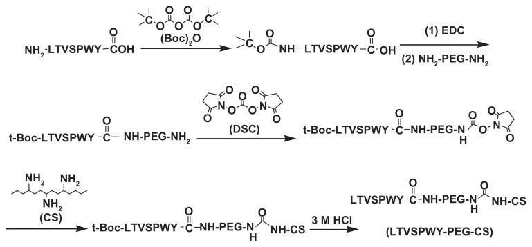
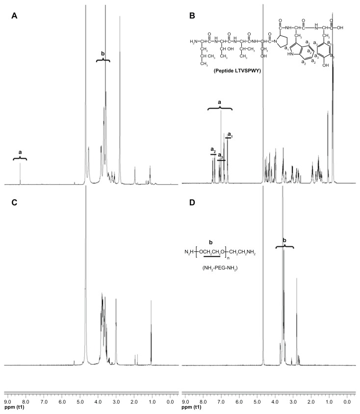
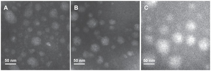
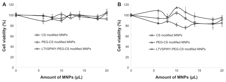
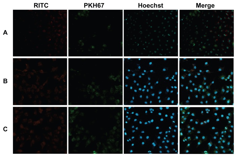
LTVSPWY peptide-modified PEGylated chitosan (LTVSPWY-PEG-CS) was synthesized by chemical reaction, and the chemical structure was confirmed by (1)H-NMR. LTVSPWY-PEG-CS-modified magnetic nanoparticles were prepared successfully using the solvent diffusion method. Their particle size, size distribution, and zeta potential were measured by dynamic light scattering and electrophoretic mobility, and their surface morphology was investigated by transmission electron microscopy. To investigate their selective targeting ability, the cellular uptake of the LTVSPWY-PEG-CS-modified magnetic nanoparticles was observed in a cocultured system of SKOV-3 cells which overexpress HER2 and A549 cells which are HER2-negative. The in vitro cytotoxicity of these nanoparticles in SKOV-3 and A549 cells was measured using the MTT method. The SKOV-3-bearing nude mouse model was used to investigate the tumor targeting ability of the magnetic nanoparticles in vivo.
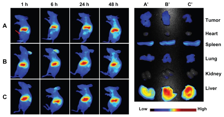
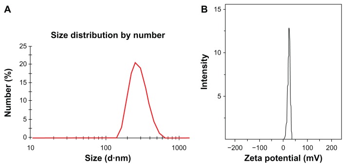
The average diameter and zeta potential of the LTVSPWY-PEG-CS-modified magnetic nanoparticles was 267.3 ± 23.4 nm and 30.5 ± 7.0 mV, respectively, with a narrow size distribution and spherical morphology. In vitro cytotoxicity tests demonstrated that these magnetic nanoparticles were carriers suitable for use in cancer diagnostics with low toxicity. With modification of the LTVSPWY homing peptide, magnetic nanoparticles could be selectively taken up by SKOV-3 cells overexpressing HER2 when cocultured with HER2-negative A549 cells. In vivo biodistribution results suggest that treatment with LTVSPWY-PEG-CS-modified magnetic nanoparticles/DiR enabled tumors to be identified and diagnosed more rapidly and efficiently in vivo.
LTVSPWY-PEG-CS-modified magnetic nanoparticles are a promising contrast agent for early detection of tumors overexpressing HER2 and further diagnostic application.
磁共振成像(MRI)作为一种诊断工具,在现代临床医学中得到了广泛应用,它提供了具有高时空分辨率的活生物体中生物现象的非侵入性和三维可视化。因此,人们对具有高效靶向能力和细胞内化能力的磁性纳米粒子作为 MRI 对比剂给予了相当大的关注,这使得能够提供更高对比度和信息更丰富的图像来检测疾病。
通过化学反应合成了 LTVSPWY 肽修饰的聚乙二醇化壳聚糖(LTVSPWY-PEG-CS),并通过(1)H-NMR 确认了其化学结构。采用溶剂扩散法成功制备了 LTVSPWY-PEG-CS 修饰的磁性纳米粒子。通过动态光散射和电泳迁移率测量了其粒径、粒径分布和 zeta 电位,并通过透射电子显微镜观察了其表面形态。为了研究其选择性靶向能力,在 SKOV-3 细胞(过表达 HER2)和 A549 细胞(HER2 阴性)的共培养体系中观察了 LTVSPWY-PEG-CS 修饰的磁性纳米粒子的细胞摄取情况。采用 MTT 法测定了这些纳米粒子在 SKOV-3 和 A549 细胞中的体外细胞毒性。采用 SKOV-3 荷瘤裸鼠模型研究了体内磁性纳米粒子的肿瘤靶向能力。
LTVSPWY-PEG-CS 修饰的磁性纳米粒子的平均直径和 zeta 电位分别为 267.3±23.4nm 和 30.5±7.0mV,粒径分布较窄,呈球形。体外细胞毒性试验表明,这些磁性纳米粒子是毒性低的适合用于癌症诊断的载体。通过修饰 LTVSPWY 归巢肽,当与 HER2 阴性的 A549 细胞共培养时,磁性纳米粒子可以被过表达 HER2 的 SKOV-3 细胞选择性摄取。体内分布结果表明,LTVSPWY-PEG-CS 修饰的磁性纳米粒子/ DiR 治疗可使肿瘤在体内更快、更有效地被识别和诊断。
LTVSPWY-PEG-CS 修饰的磁性纳米粒子是一种很有前途的用于过表达 HER2 的肿瘤早期检测和进一步诊断应用的对比剂。