Department of Biochemistry and Molecular Biology, Bio21 Molecular Science and Biotechnology Institute, The University of Melbourne, Victoria, Australia.
Diabetes. 2012 Nov;61(11):3018-25. doi: 10.2337/db11-1333. Epub 2012 Aug 7.
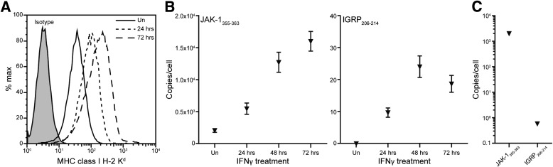
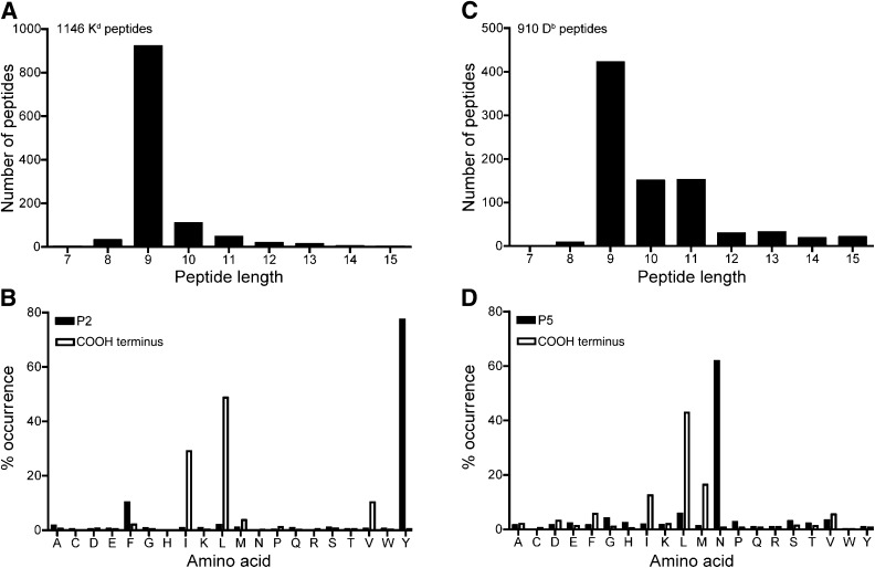
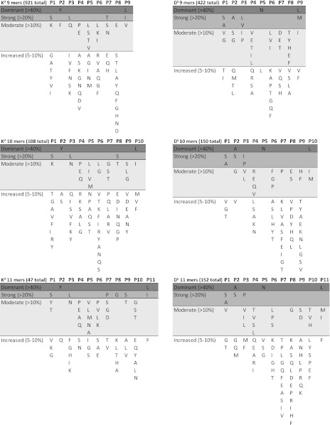
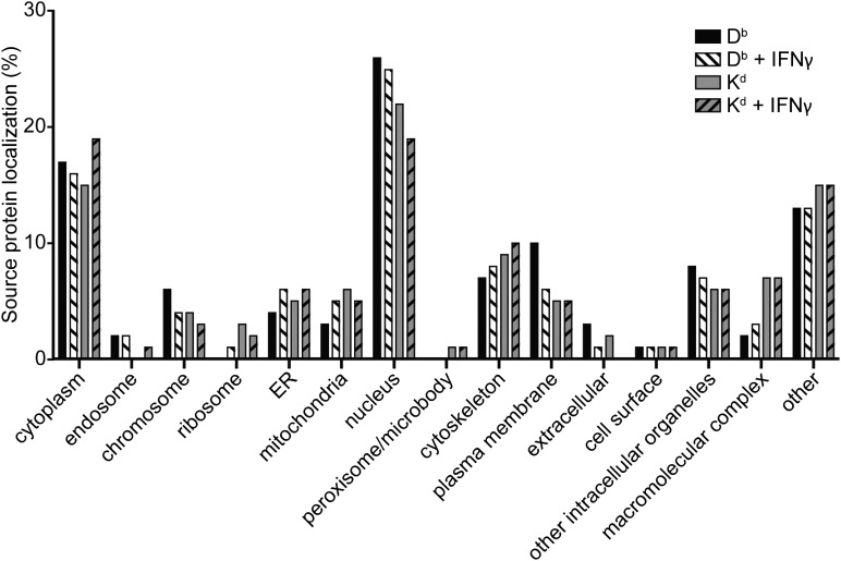
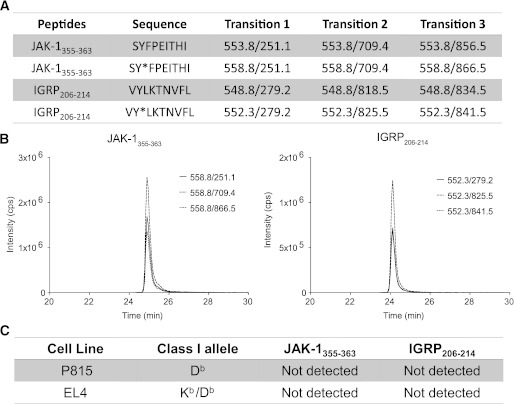
Type 1 diabetes is characterized by the autoimmune destruction of pancreatic β-cells. Recognition of major histocompatibility complex (MHC)-bound peptides is critical for both the initiation and progression of disease. In this study, MHC peptide complexes were purified from NIT-1 β-cells, interferon-γ (IFN-γ)-treated NIT-1 cells, splenic and thymic tissue of 12-week-old NOD mice, and peptides identified by mass spectrometry. In addition to global liquid chromatography-tandem mass spectrometry analysis, the targeted approach of multiple-reaction monitoring was used to quantitate the immunodominant K(d)-restricted T-cell epitope islet-specific glucose-6-phosphatase catalytic subunit-related protein (IGRP)₂₀₆₋₂₁₄. We identified >2,000 MHC-bound peptides; 1,100 of these presented by β-cells grown under normal conditions or after exposure to IFN-γ. These include sequences from a number of known autoantigens. Quantitation of IGRP₂₀₆₋₂₁₄ revealed low-level presentation by K(d) (25 complexes/cell) on NIT-1 cells after IFN-γ treatment compared with the simultaneous presentation of the endogenously processed K(d)-restricted peptide Janus kinase-1₃₅₅₋₃₆₃ (15,000 copies/cell). We have successfully sequenced peptides from NIT-1 β-cells under basal and inflammatory conditions. We have shown the feasibility of quantitating disease-associated peptides and provide the first direct demonstration of the disparity between presentation of a known autoantigenic epitope and a common endogenously presented peptide.
1 型糖尿病的特征是胰岛β细胞的自身免疫破坏。主要组织相容性复合物 (MHC) 结合肽的识别对于疾病的起始和进展都至关重要。在这项研究中,MHC 肽复合物从 NIT-1β细胞、干扰素-γ (IFN-γ) 处理的 NIT-1 细胞、12 周龄 NOD 小鼠的脾和胸腺组织中被分离出来,并通过质谱法鉴定出肽。除了全局液相色谱-串联质谱分析外,还使用多重反应监测的靶向方法来定量测定免疫优势 K(d)限制的 T 细胞表位胰岛特异性葡萄糖-6-磷酸酶催化亚基相关蛋白 (IGRP)₂₀₆₋₂₁₄。我们鉴定了 >2000 个 MHC 结合肽;其中 1100 个由β细胞在正常条件下或暴露于 IFN-γ后生长时呈递。这些包括一些已知自身抗原的序列。对 IGRP₂₀₆₋₂₁₄ 的定量分析显示,与同时呈递内源性加工的 K(d)限制肽 Janus 激酶-1₃₅₅₋₃₆₃(15000 个拷贝/细胞)相比,IFN-γ处理后的 NIT-1 细胞上 K(d)(25 个复合物/细胞)的低水平呈递。我们已经成功地在基础和炎症条件下对 NIT-1β细胞中的肽进行了测序。我们已经证明了定量测定与疾病相关的肽的可行性,并首次直接证明了已知自身抗原表位和常见内源性呈递肽之间的差异。