Department of Neuroscience, Section Medical Physiology, University Medical Center Groningen, University of Groningen, A. Deusinglaan 1, 9713 AV, Groningen, The Netherlands.
J Neuroinflammation. 2012 Aug 16;9:198. doi: 10.1186/1742-2094-9-198.
Neuroprotective and neurotrophic properties of leukemia inhibitory factor (LIF) have been widely reported. In the central nervous system (CNS), astrocytes are the major source for LIF, expression of which is enhanced following disturbances leading to neuronal damage. How astrocytic LIF expression is regulated, however, has remained an unanswered question. Since neuronal stress is associated with production of extracellular adenosine, we investigated whether LIF expression in astrocytes was mediated through adenosine receptor signaling.
Mouse cortical neuronal and astrocyte cultures from wild-type and adenosine A(2B) receptor knock-out animals, as well as adenosine receptor agonists/antagonists and various enzymatic inhibitors, were used to study LIF expression and release in astrocytes. When needed, a one-way analysis of variance (ANOVA) followed by Bonferroni post-hoc test was used for statistical analysis.
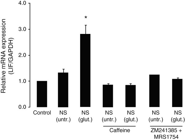
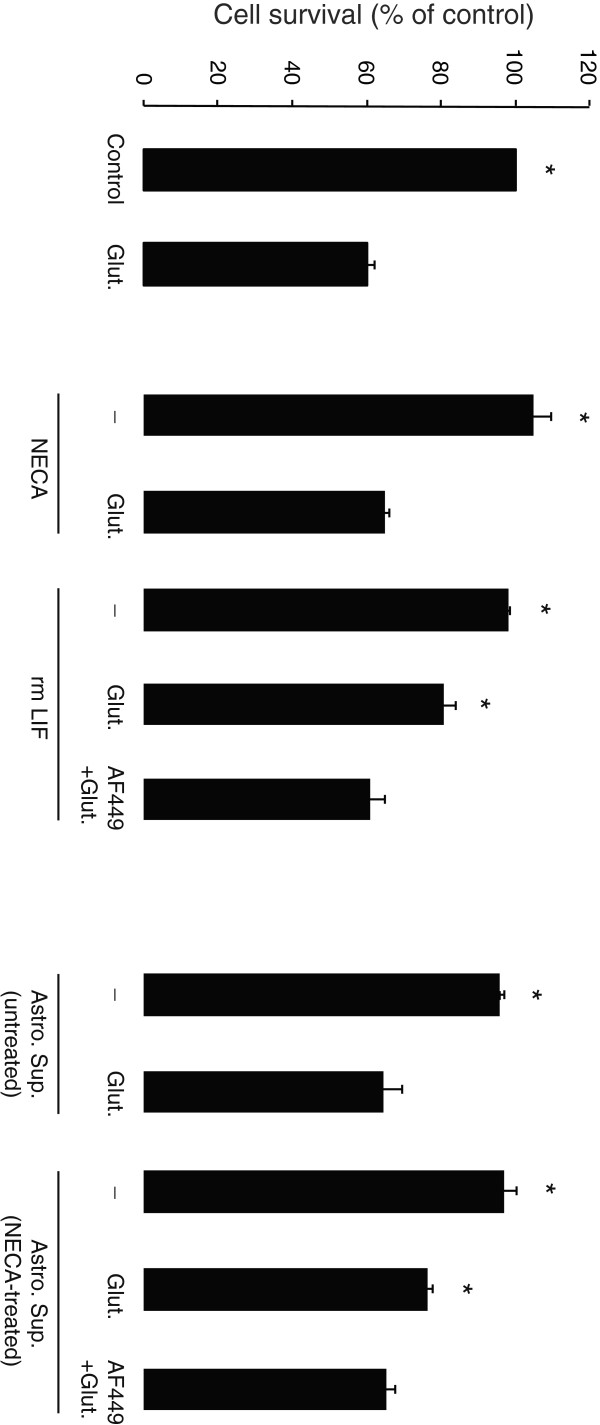
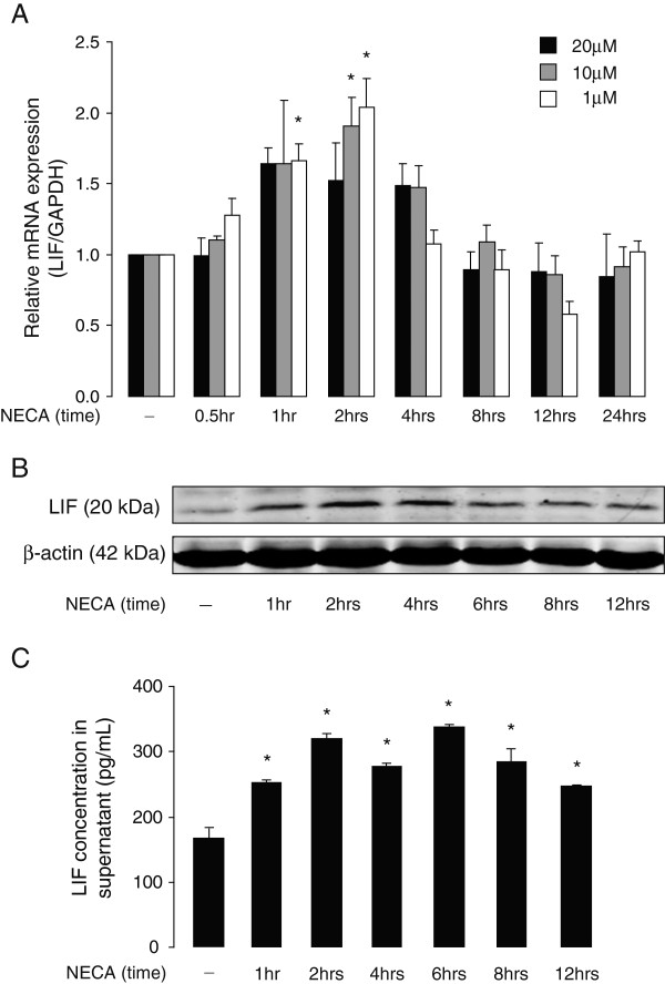
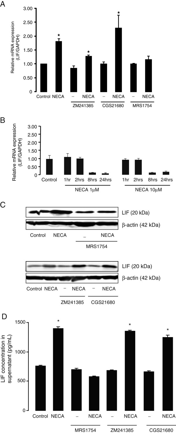
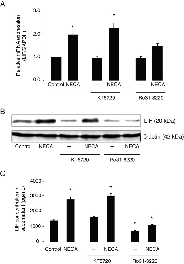
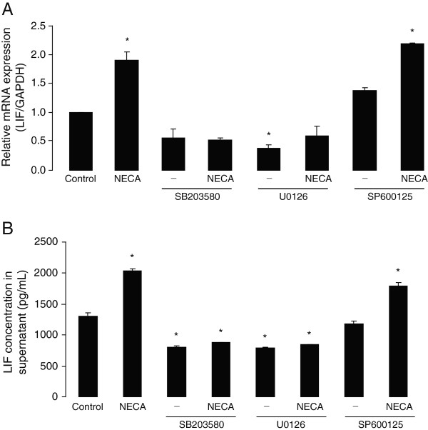
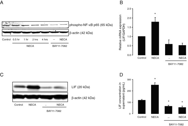
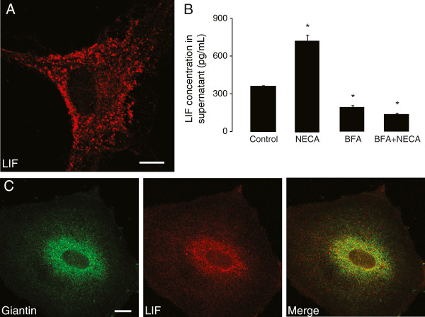
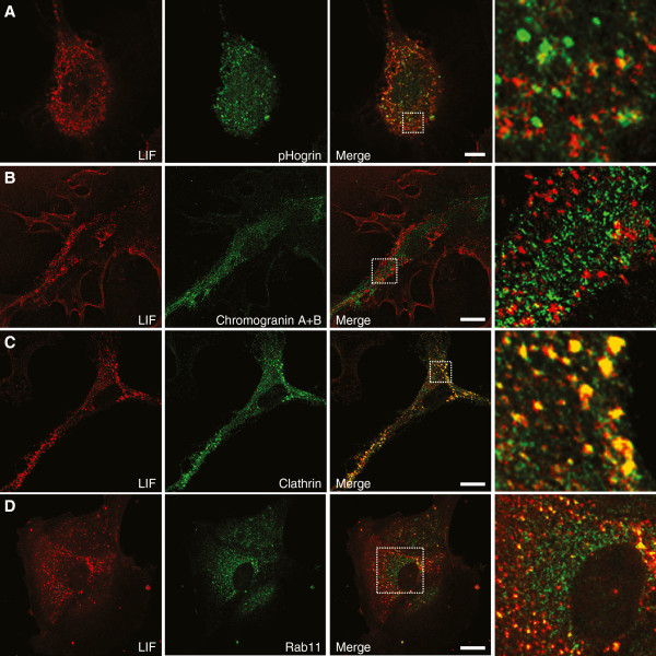
We show here that glutamate-stressed cortical neurons induce LIF expression through activation of adenosine A(2B) receptor subtype in cultured astrocytes and require signaling of protein kinase C (PKC), mitogen-activated protein kinases (MAPKs: p38 and ERK1/2), and the nuclear transcription factor (NF)-κB. Moreover, LIF concentration in the supernatant in response to 5'-N-ethylcarboxamide (NECA) stimulation was directly correlated to de novo protein synthesis, suggesting that LIF release did not occur through a regulated release pathway. Immunocytochemistry experiments show that LIF-containing vesicles co-localize with clathrin and Rab11, but not with pHogrin, Chromogranin (Cg)A and CgB, suggesting that LIF might be secreted through recycling endosomes. We further show that pre-treatment with supernatants from NECA-treated astrocytes increased survival of cultured cortical neurons against glutamate, which was absent when the supernatants were pre-treated with an anti-LIF neutralizing antibody.
Adenosine from glutamate-stressed neurons induces rapid LIF release in astrocytes. This rapid release of LIF promotes the survival of cortical neurons against excitotoxicity.
白血病抑制因子(LIF)具有神经保护和神经营养特性,这一特性已得到广泛报道。在中枢神经系统(CNS)中,星形胶质细胞是 LIF 的主要来源,在导致神经元损伤的干扰后,其表达会增强。然而,星形胶质细胞中 LIF 表达是如何调节的,仍然是一个悬而未决的问题。由于神经元应激与细胞外腺苷的产生有关,我们研究了星形胶质细胞中 LIF 的表达是否通过腺苷受体信号转导来介导。
使用来自野生型和腺苷 A(2B)受体敲除动物的皮质神经元和星形胶质细胞培养物以及腺苷受体激动剂/拮抗剂和各种酶抑制剂,研究星形胶质细胞中 LIF 的表达和释放。当需要时,采用单因素方差分析(ANOVA),然后进行 Bonferroni 事后检验进行统计学分析。
我们在这里展示,谷氨酸刺激的皮质神经元通过激活培养的星形胶质细胞中的腺苷 A(2B)受体亚型诱导 LIF 表达,并需要蛋白激酶 C(PKC)、丝裂原活化蛋白激酶(MAPKs:p38 和 ERK1/2)和核转录因子(NF)-κB 的信号转导。此外,响应 5'-N-乙基羧酰胺(NECA)刺激,上清液中 LIF 的浓度与从头蛋白质合成直接相关,这表明 LIF 的释放不是通过受调控的释放途径发生的。免疫细胞化学实验表明,含 LIF 的囊泡与网格蛋白和 Rab11 共定位,但与 pHogrin、嗜铬粒蛋白(Cg)A 和 CgB 不共定位,表明 LIF 可能通过再循环内体分泌。我们进一步表明,用 NECA 处理的星形胶质细胞上清液预处理可增加培养的皮质神经元对谷氨酸的存活,而用抗 LIF 中和抗体预处理上清液则会使存活丧失。
谷氨酸应激神经元产生的腺苷诱导星形胶质细胞中 LIF 的快速释放。这种快速释放的 LIF 促进了皮质神经元对抗兴奋性毒性的存活。