Electronic Materials Research Institute, Northeastern University, Boston MA USA.
Theranostics. 2012;2(7):714-22. doi: 10.7150/thno.3927. Epub 2012 Jul 31.
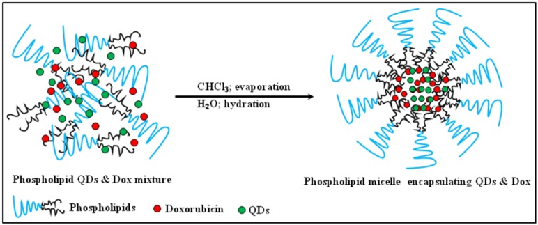
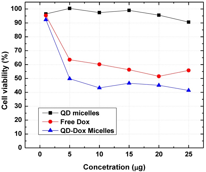
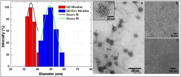
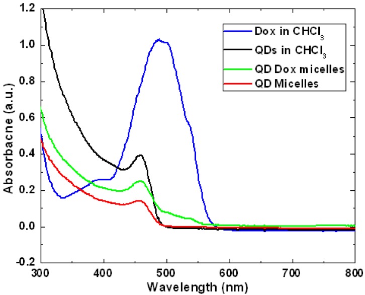
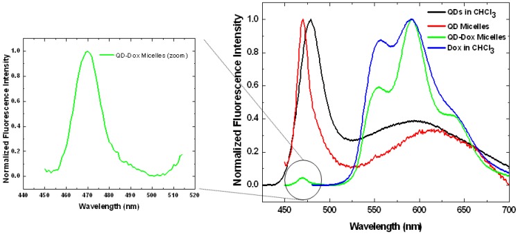
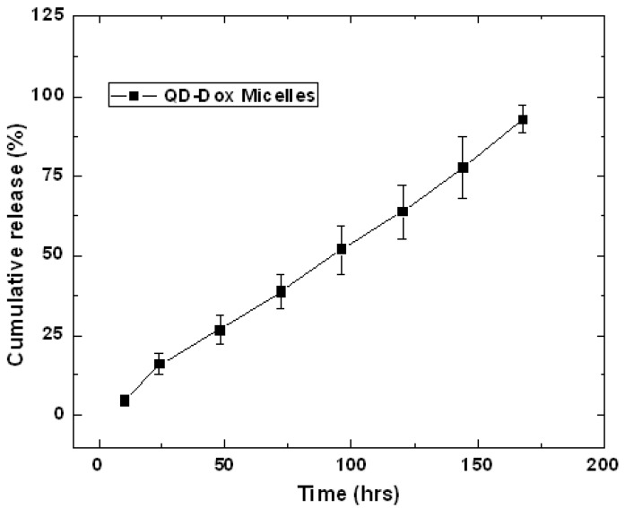
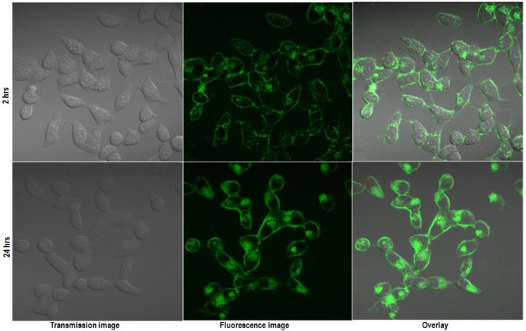
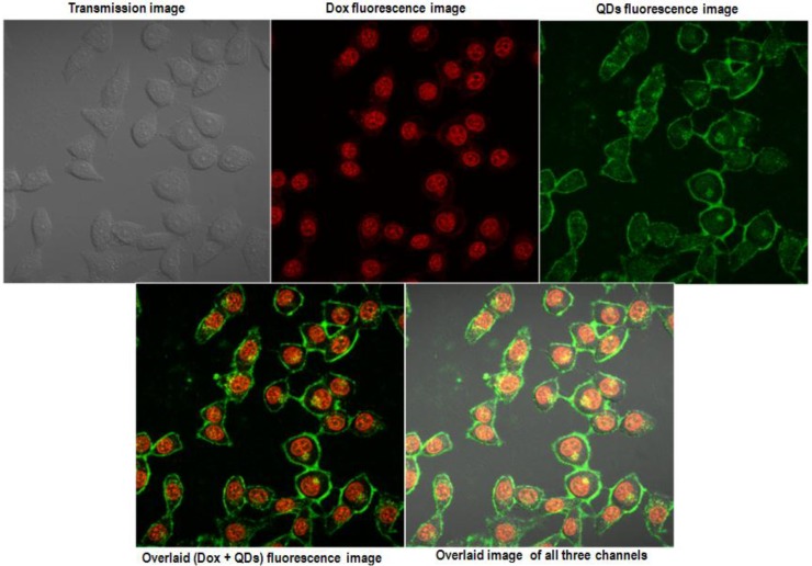
For the past decade engineered nanoplatforms have seen a momentous progress in developing a multimodal theranostic formulation which can be simultaneously used for imaging and therapy. In this report we describe the synthesis and application of theranostic phospholipid based polymeric micelles for optical fluorescence imaging and controlled drug delivery. CdSe quantum dots (QDs) and anti-cancer drug, doxorubicin (Dox), were co-encapsulated into the hydrophobic core of the micelles. The micelles are characterized using optical spectroscopy for characteristic absorbance and fluorescence features of QDs and Dox. TEM and DLS studies yielded a size of <50 nm for the micellar formulations with very narrow size distribution. A sustained release of the drug was observed from the co-encapsulated micellar formulation. In vitro optical fluorescence imaging and cytotoxicity studies with HeLa cell line demonstrated the potential of these micellar systems as efficient optical imaging and therapeutic probes.
在过去的十年中,工程纳米平台在开发可同时用于成像和治疗的多模式治疗制剂方面取得了重大进展。在本报告中,我们描述了基于治疗性磷脂的聚合物胶束的合成与应用,用于光学荧光成像和控制药物递送。将 CdSe 量子点(QD)和抗癌药物阿霉素(Dox)共同包封到胶束的疏水核中。使用光学光谱法对 QD 和 Dox 的特征吸收和荧光特性对胶束进行了表征。TEM 和 DLS 研究得到了<50nm 的胶束制剂尺寸,具有非常窄的尺寸分布。从共包封的胶束制剂中观察到药物的持续释放。用 HeLa 细胞系进行的体外光学荧光成像和细胞毒性研究表明,这些胶束系统具有作为有效光学成像和治疗探针的潜力。