• 文献检索
  • 文档翻译
  • 深度研究
  • 学术资讯
  • Suppr Zotero 插件Zotero 插件
  • 邀请有礼
  • 套餐&价格
  • 历史记录
应用&插件
Suppr Zotero 插件Zotero 插件浏览器插件Mac 客户端Windows 客户端微信小程序
定价
高级版会员购买积分包购买API积分包
服务
文献检索文档翻译深度研究API 文档MCP 服务
关于我们
关于 Suppr公司介绍联系我们用户协议隐私条款
关注我们

Suppr 超能文献

核心技术专利:CN118964589B侵权必究
粤ICP备2023148730 号-1Suppr @ 2026

文献检索

告别复杂PubMed语法,用中文像聊天一样搜索,搜遍4000万医学文献。AI智能推荐,让科研检索更轻松。

立即免费搜索

文件翻译

保留排版,准确专业,支持PDF/Word/PPT等文件格式,支持 12+语言互译。

免费翻译文档

深度研究

AI帮你快速写综述,25分钟生成高质量综述,智能提取关键信息,辅助科研写作。

立即免费体验

应用非散瞳超广角激光扫描检眼镜(Optomap)评估糖尿病性视网膜病变与 ETDRS 7 区域立体摄影比较。

Assessment of diabetic retinopathy using nonmydriatic ultra-widefield scanning laser ophthalmoscopy (Optomap) compared with ETDRS 7-field stereo photography.

机构信息

Department of Ophthalmology, Ludwig Maximilian University, Munich, Germany.

出版信息

Diabetes Care. 2012 Dec;35(12):2459-63. doi: 10.2337/dc12-0346. Epub 2012 Aug 21.

DOI:10.2337/dc12-0346
PMID:22912430
原文链接:https://pmc.ncbi.nlm.nih.gov/articles/PMC3507573/
Abstract

OBJECTIVE

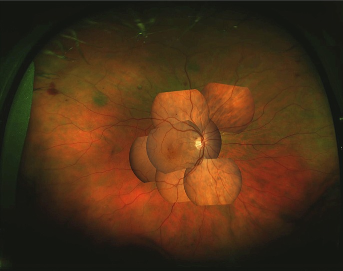
To compare the diagnostic properties of a nonmydriatic 200° ultra-widefield scanning laser ophthalmoscope (SLO) versus mydriatic Early Treatment of Diabetic Retinopathy Study (ETDRS) 7-field photography for diabetic retinopathy (DR) screening.

RESEARCH DESIGN AND METHODS

A consecutive series of 212 eyes of 141 patients with different levels of DR were examined. Grading of DR and clinically significant macular edema (CSME) from mydriatic ETDRS 7-field stereo photography was compared with grading obtained by Optomap Panoramic 200 SLO images. All SLO scans were performed through an undilated pupil, and no additional clinical information was used for evaluation of all images by the two independent, masked, expert graders.

RESULTS

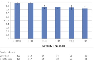
Twenty-two eyes from ETDRS 7-field photography and 12 eyes from Optomap were not gradable by at least one grader because of poor image quality. A total of 144 eyes were analyzed regarding DR level and 155 eyes regarding CSME. For ETDRS 7-field photography, 22 eyes (18 for grader 2) had no or mild DR (ETDRS levels ≤ 20) and 117 eyes (111 for grader 2) had no CSME. A highly substantial agreement between both Optomap DR and CSME grading and ETDRS 7-field photography existed with κ = 0.79 for DR and 0.73 for CSME for grader 1, and κ = 0.77 (DR) and 0.77 (CSME) for grader 2.

CONCLUSIONS

Determination of CSME and grading of DR level from Optomap Panoramic 200 nonmydriatic images show a positive correlation with mydriatic ETDRS 7-field stereo photography. Both techniques are of sufficient quality to assess DR and CSME. Optomap Panoramic 200 images cover a larger retinal area and therefore may offer additional diagnostic properties.

摘要

目的

比较非散瞳 200°超广角扫描激光检眼镜(SLO)与散瞳糖尿病眼病研究(ETDRS)7 视野摄影在糖尿病视网膜病变(DR)筛查中的诊断性能。

研究设计与方法

对 141 例不同程度 DR 的 212 只眼进行了连续系列检查。将散瞳 ETDRS 7 视野立体摄影的 DR 和临床显著黄斑水肿(CSME)分级与 Optomap Panoramic 200 SLO 图像获得的分级进行比较。所有 SLO 扫描均在未散瞳的瞳孔下进行,两名独立的、盲法、专家级评估员在评估所有图像时未使用任何其他临床信息。

结果

由于图像质量差,至少有一名评估员无法对 ETDRS 7 视野摄影的 22 只眼和 Optomap 的 12 只眼进行分级。共有 144 只眼进行了 DR 分级分析,155 只眼进行了 CSME 分级分析。对于 ETDRS 7 视野摄影,22 只眼(2 级评估员有 22 只眼)无或轻度 DR(ETDRS 分级≤20),117 只眼(2 级评估员有 111 只眼)无 CSME。Optomap 的 DR 和 CSME 分级与 ETDRS 7 视野摄影之间存在高度一致性,评估员 1 的κ值分别为 0.79(DR)和 0.73(CSME),评估员 2 的κ值分别为 0.77(DR)和 0.77(CSME)。

结论

Optomap Panoramic 200 非散瞳图像确定 CSME 和分级 DR 水平与散瞳 ETDRS 7 视野立体摄影具有正相关性。两种技术都具有足够的质量来评估 DR 和 CSME。Optomap Panoramic 200 图像覆盖了更大的视网膜区域,因此可能具有额外的诊断性能。

https://cdn.ncbi.nlm.nih.gov/pmc/blobs/f9a6/3507573/35855b7512e8/2459fig2.jpg
https://cdn.ncbi.nlm.nih.gov/pmc/blobs/f9a6/3507573/9b5af7e6dff5/2459fig1.jpg
https://cdn.ncbi.nlm.nih.gov/pmc/blobs/f9a6/3507573/35855b7512e8/2459fig2.jpg
https://cdn.ncbi.nlm.nih.gov/pmc/blobs/f9a6/3507573/9b5af7e6dff5/2459fig1.jpg
https://cdn.ncbi.nlm.nih.gov/pmc/blobs/f9a6/3507573/35855b7512e8/2459fig2.jpg

相似文献

1
Assessment of diabetic retinopathy using nonmydriatic ultra-widefield scanning laser ophthalmoscopy (Optomap) compared with ETDRS 7-field stereo photography.应用非散瞳超广角激光扫描检眼镜(Optomap)评估糖尿病性视网膜病变与 ETDRS 7 区域立体摄影比较。
Diabetes Care. 2012 Dec;35(12):2459-63. doi: 10.2337/dc12-0346. Epub 2012 Aug 21.
2
[Diabetic retinopathy: comparison of the diagnostic features of ultra-widefield scanning laser ophthalmoscopy Optomap with ETDRS 7-field fundus photography].[糖尿病视网膜病变:超广角扫描激光检眼镜Optomap与ETDRS七视野眼底摄影诊断特征的比较]
Ophthalmologe. 2011 Feb;108(2):117-23. doi: 10.1007/s00347-010-2226-4.
3
Nonmydriatic screening for diabetic retinopathy by ultra-widefield scanning laser ophthalmoscopy (Optomap).通过超广角扫描激光检眼镜(Optomap)进行糖尿病视网膜病变的免散瞳筛查。
Graefes Arch Clin Exp Ophthalmol. 2008 Feb;246(2):229-35. doi: 10.1007/s00417-007-0631-4. Epub 2007 Jul 11.
4
Nonmydriatic ultra-wide-field scanning laser ophthalmoscopy (Optomap) versus two-field fundus photography in diabetic retinopathy.非散瞳超广角激光扫描检眼镜(Optomap)与两种眼底照相在糖尿病视网膜病变中的比较。
Ophthalmologica. 2014;231(1):31-6. doi: 10.1159/000355092. Epub 2013 Nov 13.
5
Comparison between Early Treatment Diabetic Retinopathy Study 7-field retinal photos and non-mydriatic, mydriatic and mydriatic steered widefield scanning laser ophthalmoscopy for assessment of diabetic retinopathy.早期治疗糖尿病性视网膜病变研究的7视野视网膜照片与非散瞳、散瞳及散瞳引导的超广角扫描激光检眼镜在糖尿病性视网膜病变评估中的比较。
J Diabetes Complications. 2015 Jan-Feb;29(1):99-104. doi: 10.1016/j.jdiacomp.2014.08.009. Epub 2014 Aug 28.
6
Nonmydriatic ultrawide field retinal imaging compared with dilated standard 7-field 35-mm photography and retinal specialist examination for evaluation of diabetic retinopathy.非散瞳超广角视网膜成像与散瞳标准 7 区域 35mm 摄影及视网膜专科医师检查在糖尿病视网膜病变评估中的比较。
Am J Ophthalmol. 2012 Sep;154(3):549-559.e2. doi: 10.1016/j.ajo.2012.03.019. Epub 2012 May 23.
7
Comparison of Widefield Laser Ophthalmoscopy and ETDRS Retinal Area for Diabetic Retinopathy.广角激光眼底镜检查与糖尿病视网膜病变早期治疗糖尿病性视网膜病变研究(ETDRS)视网膜区域的比较
Ophthalmol Sci. 2022 Jun 28;2(4):100190. doi: 10.1016/j.xops.2022.100190. eCollection 2022 Dec.
8
Web-based grading of compressed stereoscopic digital photography versus standard slide film photography for the diagnosis of diabetic retinopathy.基于网络的压缩立体数码摄影与标准幻灯片胶片摄影在糖尿病视网膜病变诊断中的分级比较
Ophthalmology. 2007 Sep;114(9):1748-54. doi: 10.1016/j.ophtha.2006.12.010. Epub 2007 Mar 21.
9
ETDRS grading with CLARUS ultra-widefield images shows agreement with 7-fields colour fundus photography.ETDRS 分级与 CLARUS 超广角图像具有一致性,与 7 区域彩色眼底照相一致。
BMC Ophthalmol. 2024 Sep 3;24(1):387. doi: 10.1186/s12886-024-03537-z.
10
The sensitivity and specificity of single-field nonmydriatic monochromatic digital fundus photography with remote image interpretation for diabetic retinopathy screening: a comparison with ophthalmoscopy and standardized mydriatic color photography.单视野非散瞳单色数字眼底摄影结合远程图像解读用于糖尿病视网膜病变筛查的敏感性和特异性:与检眼镜检查及标准化散瞳彩色摄影的比较
Am J Ophthalmol. 2002 Aug;134(2):204-13. doi: 10.1016/s0002-9394(02)01522-2.

引用本文的文献

1
The Influence of Ultra-Wide field Fluorescein Angiography on the Diagnosis and Management of Diabetic Retinopathy.超广角荧光素血管造影对糖尿病视网膜病变诊断和治疗的影响
J Vitreoretin Dis. 2025 Aug 1:24741264251359907. doi: 10.1177/24741264251359907.
2
Quantitative Biomarkers of Diabetic Retinopathy Using Ultra-Widefield Fluorescein Angiography.利用超广角荧光素血管造影术检测糖尿病视网膜病变的定量生物标志物
Clin Ophthalmol. 2024 Jul 8;18:1961-1970. doi: 10.2147/OPTH.S462223. eCollection 2024.
3
Retinal vascular morphological characteristics in diabetic retinopathy: an artificial intelligence study using a transfer learning system to analyze ultra-wide field images.

本文引用的文献

1
Teleophthalmology assessment of diabetic retinopathy fundus images: smartphone versus standard office computer workstation.智能手机与标准办公电脑工作站在糖尿病视网膜病变眼底图像远程眼科评估中的应用比较。
Telemed J E Health. 2012 Mar;18(2):158-62. doi: 10.1089/tmj.2011.0089. Epub 2012 Feb 3.
2
Development of a mobile phone based ophthalmoscope for telemedicine.用于远程医疗的基于手机的检眼镜的开发。
Annu Int Conf IEEE Eng Med Biol Soc. 2011;2011:5236-9. doi: 10.1109/IEMBS.2011.6091295.
3
Light and portable novel device for diabetic retinopathy screening.
糖尿病视网膜病变中的视网膜血管形态特征:一项使用迁移学习系统分析超广角图像的人工智能研究。
Int J Ophthalmol. 2024 Jun 18;17(6):1001-1006. doi: 10.18240/ijo.2024.06.03. eCollection 2024.
4
Quantifying image quality in AOSLO images of photoreceptors.量化光感受器AOSLO图像的图像质量。
Biomed Opt Express. 2024 Apr 4;15(5):2849-2862. doi: 10.1364/BOE.516477. eCollection 2024 May 1.
5
American Society of Retina Specialists Clinical Practice Guidelines on Multimodal Imaging for Retinal Disease.美国视网膜专家协会关于视网膜疾病多模态成像的临床实践指南。
J Vitreoretin Dis. 2024 Mar 21;8(3):234-246. doi: 10.1177/24741264241237012. eCollection 2024 May-Jun.
6
Algorithm of automatic identification of diabetic retinopathy foci based on ultra-widefield scanning laser ophthalmoscopy.基于超广角扫描激光检眼镜的糖尿病视网膜病变病灶自动识别算法
Int J Ophthalmol. 2024 Apr 18;17(4):610-615. doi: 10.18240/ijo.2024.04.02. eCollection 2024.
7
Imaging Modalities for Assessing the Vascular Component of Diabetic Retinal Disease: Review and Consensus for an Updated Staging System.评估糖尿病视网膜病变血管成分的成像方式:更新分期系统的综述与共识
Ophthalmol Sci. 2023 Dec 10;4(3):100449. doi: 10.1016/j.xops.2023.100449. eCollection 2024 May-Jun.
8
Present and future screening programs for diabetic retinopathy: a narrative review.糖尿病视网膜病变的当前及未来筛查项目:一篇叙述性综述
Int J Retina Vitreous. 2024 Feb 3;10(1):14. doi: 10.1186/s40942-024-00534-8.
9
Wide-field imaging with smartphone based fundus camera: grading of severity of diabetic retinopathy and locating peripheral lesions in diabetic retinopathy.基于智能手机的眼底相机广角成像:糖尿病视网膜病变严重程度分级和糖尿病视网膜病变周边病变定位。
Eye (Lond). 2024 Jun;38(8):1471-1476. doi: 10.1038/s41433-024-02928-2. Epub 2024 Jan 31.
10
Ultra-Widefield Imaging as a Teleophthalmology Screening Tool for Ocular Pathology.超广角成像作为眼部病理学的远程眼科筛查工具
Clin Ophthalmol. 2023 Oct 30;17:3225-3234. doi: 10.2147/OPTH.S433864. eCollection 2023.
用于糖尿病视网膜病变筛查的轻巧便携新型设备。
Clin Exp Ophthalmol. 2012 Jan-Feb;40(1):e40-6. doi: 10.1111/j.1442-9071.2011.02732.x. Epub 2011 Dec 23.
4
Improving diabetic retinopathy screening through a statewide telemedicine program at a large federally qualified health center.通过一家大型联邦合格健康中心的全州远程医疗项目改善糖尿病视网膜病变筛查。
J Health Care Poor Underserved. 2011 Aug;22(3):804-16. doi: 10.1353/hpu.2011.0066.
5
Imaging of diabetic retinopathy and diabetic macular edema.糖尿病视网膜病变与糖尿病黄斑水肿的影像学表现
Curr Diab Rep. 2011 Aug;11(4):236-43. doi: 10.1007/s11892-011-0203-1.
6
[Diabetic retinopathy: comparison of the diagnostic features of ultra-widefield scanning laser ophthalmoscopy Optomap with ETDRS 7-field fundus photography].[糖尿病视网膜病变:超广角扫描激光检眼镜Optomap与ETDRS七视野眼底摄影诊断特征的比较]
Ophthalmologe. 2011 Feb;108(2):117-23. doi: 10.1007/s00347-010-2226-4.
7
Images in cardiovascular medicine. Wide-field scanning laser ophthalmoscope imaging and angiography of central retinal vein occlusion.心血管医学影像。视网膜中央静脉阻塞的广角扫描激光检眼镜成像及血管造影
Circulation. 2010 Mar 30;121(12):1459-60. doi: 10.1161/CIR.0b013e3181d99c22.
8
Diabetic macular oedema: physical, physiological and molecular factors contribute to this pathological process.糖尿病性黄斑水肿:物理、生理和分子因素共同导致这一病理过程。
Acta Ophthalmol. 2010 May;88(3):279-91. doi: 10.1111/j.1755-3768.2008.01501.x. Epub 2010 Mar 11.
9
Screening of diabetic retinopathy: effect of field number and mydriasis on sensitivity and specificity of digital fundus photography.糖尿病视网膜病变的筛查:视野数量和散瞳对数字眼底摄影敏感性和特异性的影响。
Diabetes Metab. 2008 Jun;34(3):290-3. doi: 10.1016/j.diabet.2007.12.007. Epub 2008 Apr 10.
10
Digital fundus image grading with the non-mydriatic Visucam(PRO NM) versus the FF450(plus) camera in diabetic retinopathy.使用非散瞳Visucam(PRO NM)与FF450(plus)相机对糖尿病视网膜病变进行数字眼底图像分级
Acta Ophthalmol. 2008 Mar;86(2):177-82. doi: 10.1111/j.1600-0420.2007.01029.x. Epub 2007 Oct 18.