Bahrami S Bahram, Veiseh Mandana, Boudreau Nancy J
Department of Surgery, University of California San Francisco, San Francisco, CA, USA.
Methods Mol Biol. 2012;916:81-96. doi: 10.1007/978-1-61779-980-8_7.
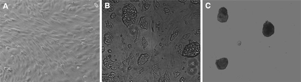
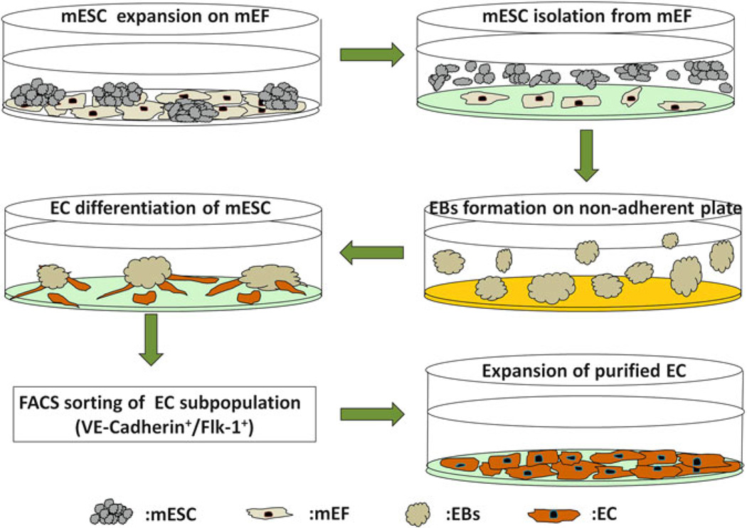
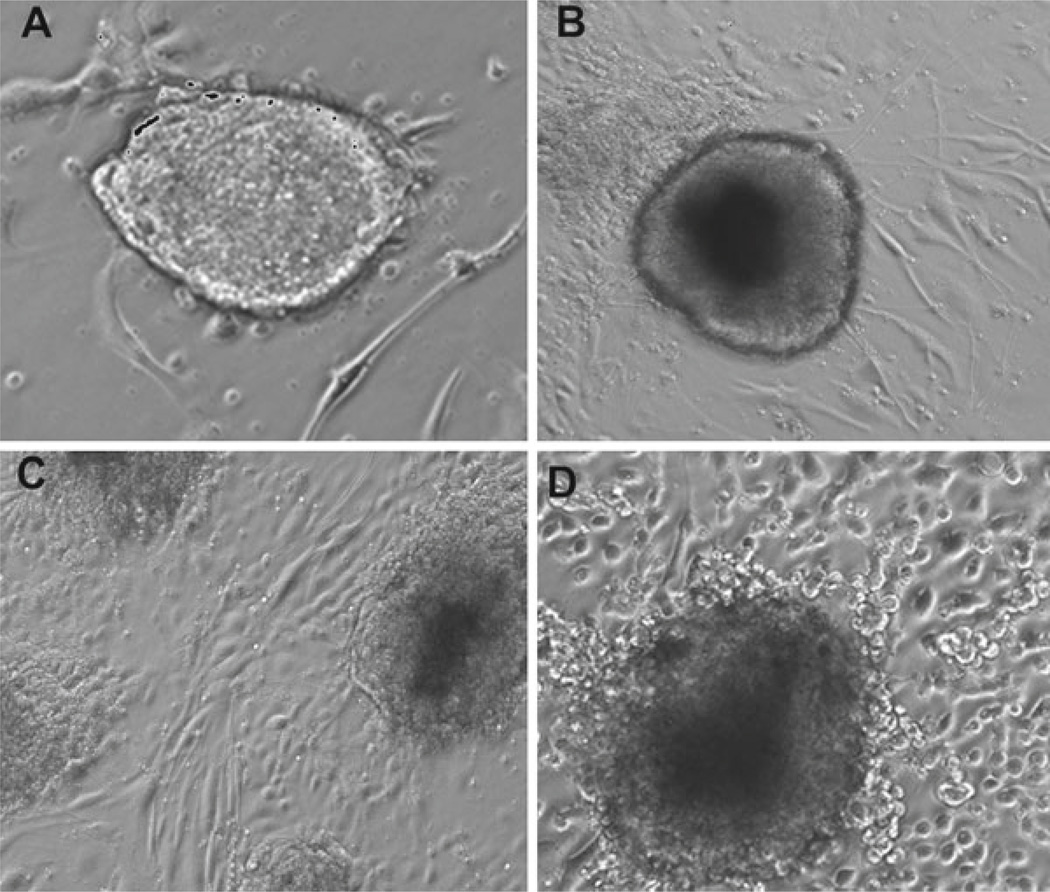
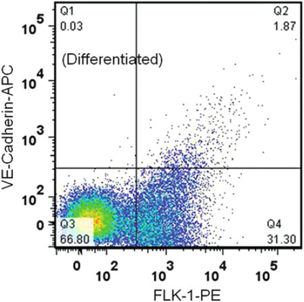
The unlimited differentiation and proliferation capacity of embryonic stem cells represents a great resource for regenerative medicine. Here, we describe a method for differentiating, isolating, and expanding endothelial cells (ECs) from mouse embryonic stem cells (mESCs). First, mESCs are expanded on a mouse embryonic fibroblast (mEF) feeder layer and partially differentiated into embryoid bodies (EBs) by growing the cells in an ultra-low attachment plate for up to 5 days. The EBs are then differentiated along the endothelial lineage using endothelial growth medium supplemented with 40 ng/mL vascular endothelial growth factor (VEGF). The differentiated endothelial population expresses both Fetal Liver Kinase 1 (Flk-1) and VE-Cadherin on the cell surface which can be further purified using a fluorescence-activated cell sorting (FACS) system and subsequently expanded on 0.1 % gelatin-coated plates. The differentiated cells can be analyzed by real-time PCR and flow cytometry to confirm enrichment of EC-specific genes and proteins.
胚胎干细胞无限的分化和增殖能力为再生医学提供了巨大的资源。在此,我们描述了一种从小鼠胚胎干细胞(mESCs)中分化、分离和扩增内皮细胞(ECs)的方法。首先,将mESCs在小鼠胚胎成纤维细胞(mEF)饲养层上扩增,并通过在超低附着板中培养细胞长达5天,使其部分分化为胚状体(EBs)。然后,使用补充有40 ng/mL血管内皮生长因子(VEGF)的内皮生长培养基,使EBs沿内皮谱系分化。分化的内皮细胞群体在细胞表面同时表达胎儿肝激酶1(Flk-1)和VE-钙黏蛋白,可使用荧光激活细胞分选(FACS)系统进一步纯化,随后在涂有0.1%明胶的培养板上扩增。分化的细胞可通过实时PCR和流式细胞术进行分析,以确认EC特异性基因和蛋白质的富集。