• 文献检索
  • 文档翻译
  • 深度研究
  • 学术资讯
  • Suppr Zotero 插件Zotero 插件
  • 邀请有礼
  • 套餐&价格
  • 历史记录
应用&插件
Suppr Zotero 插件Zotero 插件浏览器插件Mac 客户端Windows 客户端微信小程序
定价
高级版会员购买积分包购买API积分包
服务
文献检索文档翻译深度研究API 文档MCP 服务
关于我们
关于 Suppr公司介绍联系我们用户协议隐私条款
关注我们

Suppr 超能文献

核心技术专利:CN118964589B侵权必究
粤ICP备2023148730 号-1Suppr @ 2026

文献检索

告别复杂PubMed语法,用中文像聊天一样搜索,搜遍4000万医学文献。AI智能推荐,让科研检索更轻松。

立即免费搜索

文件翻译

保留排版,准确专业,支持PDF/Word/PPT等文件格式,支持 12+语言互译。

免费翻译文档

深度研究

AI帮你快速写综述,25分钟生成高质量综述,智能提取关键信息,辅助科研写作。

立即免费体验

QD-转铁蛋白生物缀合物对体外原始卵泡的入侵和生殖毒性。

The Invasion and Reproductive Toxicity of QDs-Transferrin Bioconjugates on Preantral Follicle in vitro.

机构信息

1. College of Optoelectronic Engineering, Key Laboratory of Optoelectronics Devices and Systems of Ministry of Education/Guangdong Province, Shenzhen University, Shenzhen 518060, P. R. China.

出版信息

Theranostics. 2012;2(7):734-45. doi: 10.7150/thno.4290. Epub 2012 Aug 1.

DOI:10.7150/thno.4290
PMID:22916073
原文链接:https://pmc.ncbi.nlm.nih.gov/articles/PMC3425092/
Abstract

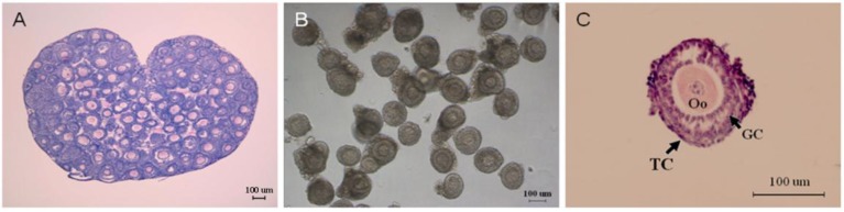
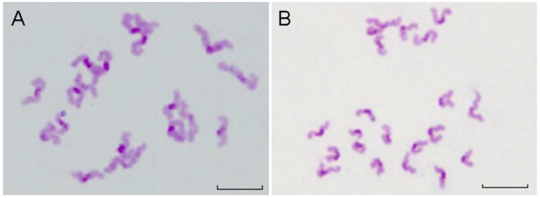
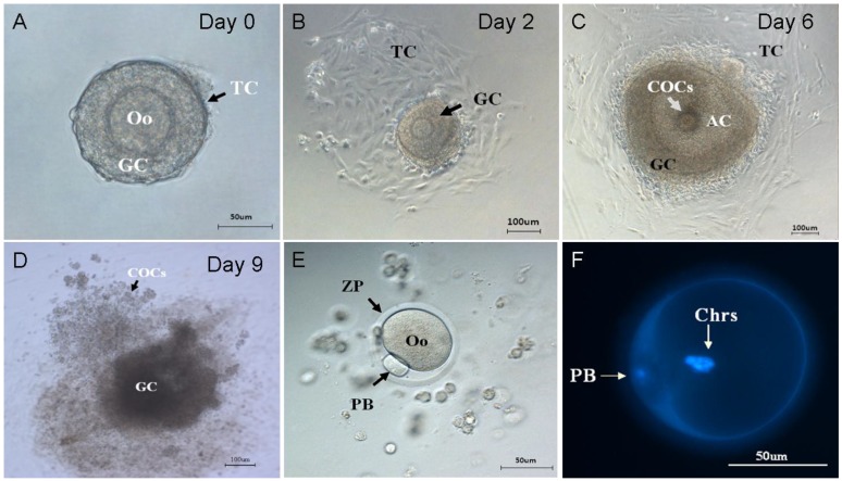
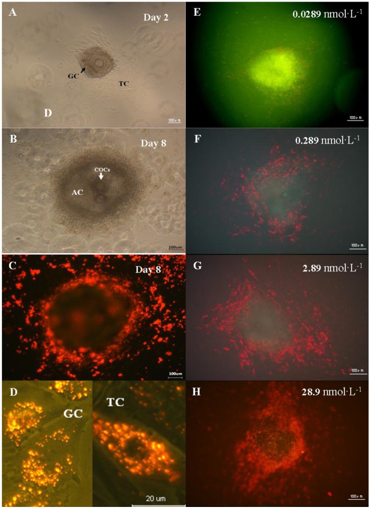
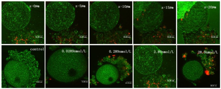
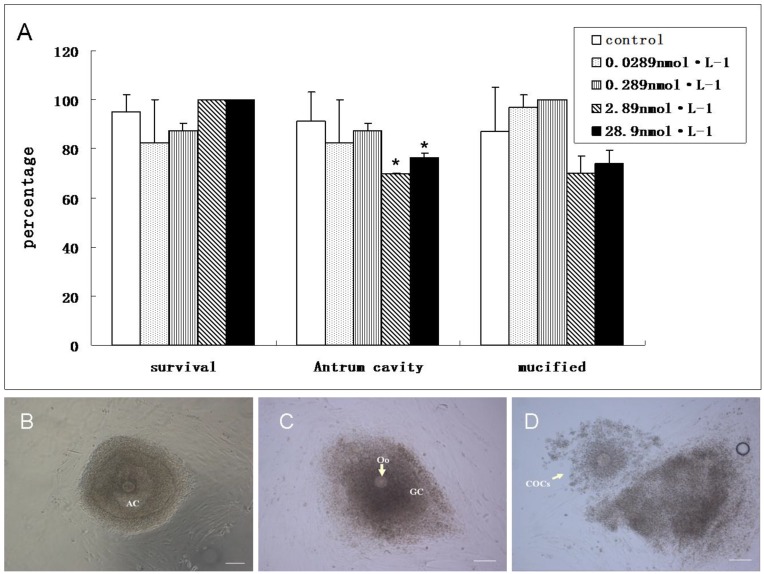
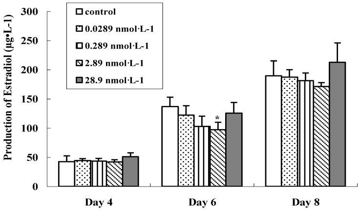
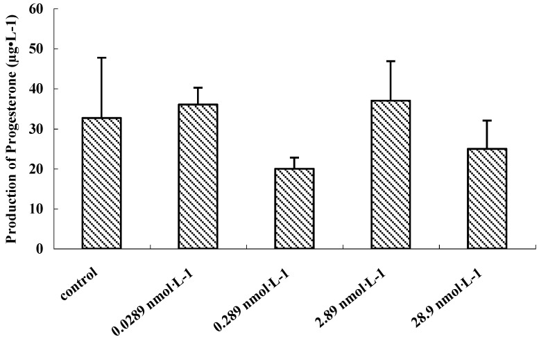
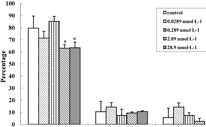
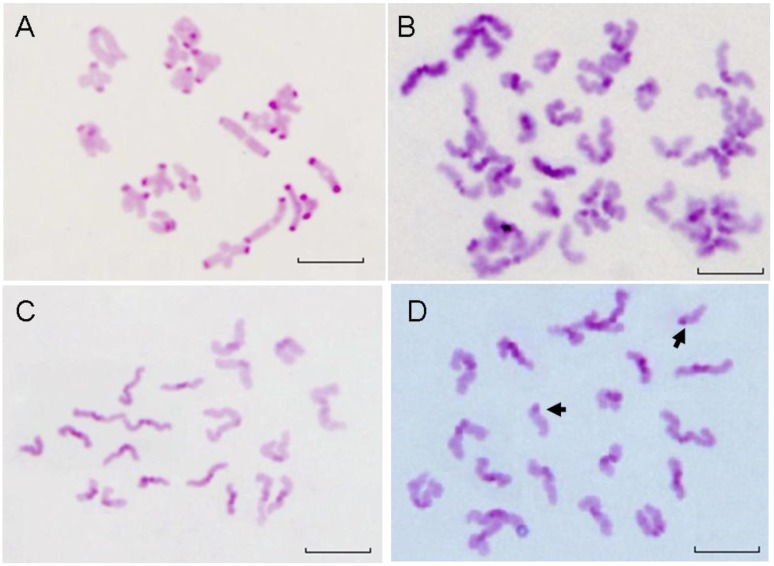
The toxicity of QD has been extensively studied over the past decade. However, the potential toxicity of QDs impedes its use for clinical research. In this work, we established a preantral follicle in vitro culture system to investigate the effects of QD-Transferrin (QDs-Tf) bioconjugates on follicle development and oocyte maturation. The preantral follicles were cultured and exposed to CdTe/ZnTe QDs-Tf bioconjugates with various concentrations and the reproductive toxicity was assessed at different time points post-treatment. The invasion of QDs-Tf for oocytes was verified by laser scanning confocal microscope. Steroid production was evaluated by immunoassay. C-band Giemsa staining was performed to observe the chromosome abnormality of oocytes. The results showed that the QDs-Tf bioconjugates could permeate into granulosa cells and theca cells, but not into oocyte. There are no obvious changes of oocyte diameter, the mucification of cumulus-oocyte-complexes and the occurrence of aneulpoidy as compared with the control group. However, delay in the antrum formation and decrease in the ratio of oocytes with first polar body were observed in QDs-Tf-treated groups. The matured oocytes with first polar body decreased significantly by ~16% (from 79.6±10 % to 63±2.9 %) when the concentration of QDs-Tf bioconjugates exceeded 2.89 nmol·L(-1) (P < 0.05). Our results implied that the CdTe/ZnTe QDs-Tf bioconjugates were reproductive toxic for follicle development, and thus also revealed that this in vitro culture system of preantral follicle is a highly sensitive tool for study on the reproductive toxicity of nanoparticles.

摘要

过去十年中,人们对 QD 的毒性进行了广泛的研究。然而,QD 的潜在毒性阻碍了其在临床研究中的应用。在这项工作中,我们建立了一个原始卵泡体外培养系统,以研究 QD-转铁蛋白(QDs-Tf)结合物对卵泡发育和卵母细胞成熟的影响。将原始卵泡进行培养并暴露于不同浓度的 CdTe/ZnTe QDs-Tf 结合物中,在处理后不同时间点评估其生殖毒性。通过激光扫描共聚焦显微镜验证 QDs-Tf 对卵母细胞的侵袭。通过免疫测定评估类固醇的产生。通过 C 带吉姆萨染色观察卵母细胞的染色体异常。结果表明,QD-Tf 结合物可以渗透到颗粒细胞和膜细胞中,但不能渗透到卵母细胞中。与对照组相比,卵母细胞直径、卵丘-卵母细胞复合体的粘液化和非整倍体的发生没有明显变化。然而,在 QDs-Tf 处理组中观察到腔形成延迟和具有第一极体的卵母细胞比例降低。当 QDs-Tf 结合物的浓度超过 2.89 nmol·L(-1)时,具有第一极体的成熟卵母细胞显著减少约 16%(从 79.6±10%降至 63±2.9%)(P<0.05)。我们的结果表明,CdTe/ZnTe QDs-Tf 结合物对卵泡发育具有生殖毒性,这也表明原始卵泡的体外培养系统是研究纳米颗粒生殖毒性的一种高度敏感工具。

https://cdn.ncbi.nlm.nih.gov/pmc/blobs/a219/3425092/10d3a45fc6a5/thnov02p0734g10.jpg
https://cdn.ncbi.nlm.nih.gov/pmc/blobs/a219/3425092/3a9ecbd4f61f/thnov02p0734g01.jpg
https://cdn.ncbi.nlm.nih.gov/pmc/blobs/a219/3425092/937bb2a0db9f/thnov02p0734g02.jpg
https://cdn.ncbi.nlm.nih.gov/pmc/blobs/a219/3425092/a3cc847a0ddd/thnov02p0734g03.jpg
https://cdn.ncbi.nlm.nih.gov/pmc/blobs/a219/3425092/e917722e74d4/thnov02p0734g04.jpg
https://cdn.ncbi.nlm.nih.gov/pmc/blobs/a219/3425092/a05410df3958/thnov02p0734g05.jpg
https://cdn.ncbi.nlm.nih.gov/pmc/blobs/a219/3425092/7130d317da0c/thnov02p0734g06.jpg
https://cdn.ncbi.nlm.nih.gov/pmc/blobs/a219/3425092/d6f4bdc0fbfb/thnov02p0734g07.jpg
https://cdn.ncbi.nlm.nih.gov/pmc/blobs/a219/3425092/786257a90475/thnov02p0734g08.jpg
https://cdn.ncbi.nlm.nih.gov/pmc/blobs/a219/3425092/2b856eb3554d/thnov02p0734g09.jpg
https://cdn.ncbi.nlm.nih.gov/pmc/blobs/a219/3425092/10d3a45fc6a5/thnov02p0734g10.jpg

相似文献

1
The Invasion and Reproductive Toxicity of QDs-Transferrin Bioconjugates on Preantral Follicle in vitro.QD-转铁蛋白生物缀合物对体外原始卵泡的入侵和生殖毒性。
Theranostics. 2012;2(7):734-45. doi: 10.7150/thno.4290. Epub 2012 Aug 1.
2
[Establishment of in-vitro mouse preantral follicle culture systems].[体外小鼠窦前卵泡培养系统的建立]
Wei Sheng Yan Jiu. 2009 Mar;38(2):153-8.
3
Effect of heat stress on the survival and development of in vitro cultured bovine preantral follicles and on in vitro maturation of cumulus-oocyte complex.热应激对体外培养的牛腔前卵泡存活与发育以及卵丘-卵母细胞复合体体外成熟的影响。
Theriogenology. 2016 Sep 1;86(4):994-1003. doi: 10.1016/j.theriogenology.2016.03.027. Epub 2016 Mar 25.
4
Effects of aromatase inhibition on in vitro follicle and oocyte development analyzed by early preantral mouse follicle culture.通过早期窦前小鼠卵泡培养分析芳香化酶抑制对体外卵泡和卵母细胞发育的影响。
Mol Reprod Dev. 2002 Apr;61(4):549-59. doi: 10.1002/mrd.10107.
5
Growth and antrum formation of bovine preantral follicles in long-term culture in vitro.牛腔前卵泡在体外长期培养中的生长及腔形成
Biol Reprod. 2000 May;62(5):1322-8. doi: 10.1095/biolreprod62.5.1322.
6
Effect of activin A and insulin-like growth factor-I on in vitro development of preantral follicles isolated from cryopreserved ovarian tissues in the mouse.激活素A和胰岛素样生长因子-I对从小鼠冷冻保存的卵巢组织中分离的腔前卵泡体外发育的影响。
Cryobiology. 2008 Dec;57(3):209-15. doi: 10.1016/j.cryobiol.2008.08.004. Epub 2008 Aug 26.
7
Effects of culture medium, serum type, and various concentrations of follicle-stimulating hormone on porcine preantral follicular development and antrum formation in vitro.培养基、血清类型及不同浓度促卵泡激素对猪腔前卵泡体外发育及窦腔形成的影响
Biol Reprod. 2002 Oct;67(4):1197-203. doi: 10.1095/biolreprod67.4.1197.
8
In vitro growth, maturation, fertilization, and embryonic development of oocytes from porcine preantral follicles.猪腔前卵泡卵母细胞的体外生长、成熟、受精及胚胎发育
Biol Reprod. 2001 Jan;64(1):375-81. doi: 10.1095/biolreprod64.1.375.
9
In vitro culture of isolated preantral and antral follicles of goats using human recombinant FSH: Concentration-dependent and stage-specific effect.使用人重组促卵泡激素对山羊分离的窦前卵泡和窦卵泡进行体外培养:浓度依赖性和阶段特异性效应。
Anim Reprod Sci. 2018 Sep;196:120-129. doi: 10.1016/j.anireprosci.2018.07.004. Epub 2018 Jul 17.
10
Activin promotes oocyte development in ovine preantral follicles in vitro.激活素在体外促进绵羊腔前卵泡中的卵母细胞发育。
Reprod Biol Endocrinol. 2003 Nov 3;1:76. doi: 10.1186/1477-7827-1-76.

引用本文的文献

1
Current Advances in the Biomedical Applications of Quantum Dots: Promises and Challenges.当前量子点在生物医学应用中的进展:前景与挑战。
Int J Mol Sci. 2023 Aug 11;24(16):12682. doi: 10.3390/ijms241612682.
2
Red CdSe/ZnS QDs' Intracellular Trafficking and Its Impact on Yeast Polarization and Actin Filament.红色 CdSe/ZnS QDs 的细胞内转位及其对酵母极性和肌动蛋白丝的影响。
Cells. 2023 Feb 2;12(3):484. doi: 10.3390/cells12030484.
3
In vitro growth (IVG) of human ovarian follicles in frozen thawed ovarian cortex tissue culture supplemented with follicular fluid under hypoxic conditions.

本文引用的文献

1
Toxicity of gold nanoparticles on somatic and reproductive cells.金纳米粒子对体细胞和生殖细胞的毒性。
Adv Exp Med Biol. 2012;733:125-33. doi: 10.1007/978-94-007-2555-3_12.
2
Folic acid modified gelatine coated quantum dots as potential reagents for in vitro cancer diagnostics.叶酸修饰的明胶涂层量子点作为体外癌症诊断的潜在试剂。
J Nanobiotechnology. 2011 Nov 10;9:50. doi: 10.1186/1477-3155-9-50.
3
Integrin targeting for tumor optical imaging.整合素靶向用于肿瘤光学成像。
在缺氧条件下,用人卵巢皮质组织冻融后培养液培养补充卵泡液,进行人卵巢卵泡的体外生长(IVG)。
Arch Gynecol Obstet. 2022 Oct;306(4):1299-1311. doi: 10.1007/s00404-022-06672-4. Epub 2022 Jul 24.
4
Eugenol Improves Follicular Survival and Development During Culture of Goat Ovarian Tissue.丁香酚可提高山羊卵巢组织培养过程中卵泡的存活率和发育能力。
Front Vet Sci. 2022 Apr 28;9:822367. doi: 10.3389/fvets.2022.822367. eCollection 2022.
5
Relative expression of microRNAs, apoptosis, and ultrastructure anomalies induced by gold nanoparticles in Trachyderma hispida (Coleoptera: Tenebrionidae).金纳米颗粒诱导糙皮侧耳蕈甲(鞘翅目:拟步甲科)中 microRNAs、细胞凋亡及超微结构异常的相对表达。
PLoS One. 2020 Nov 6;15(11):e0241837. doi: 10.1371/journal.pone.0241837. eCollection 2020.
6
Nanoparticles and female reproductive system: how do nanoparticles affect oogenesis and embryonic development.纳米颗粒与女性生殖系统:纳米颗粒如何影响卵子发生和胚胎发育。
Oncotarget. 2017 Jul 7;8(65):109799-109817. doi: 10.18632/oncotarget.19087. eCollection 2017 Dec 12.
7
The Reproductive Toxicity of CdSe/ZnS Quantum Dots on the in vivo Ovarian Function and in vitro Fertilization.CdSe/ZnS 量子点对体内卵巢功能和体外受精的生殖毒性。
Sci Rep. 2016 Nov 23;6:37677. doi: 10.1038/srep37677.
8
Engineered nanomaterials: an emerging class of novel endocrine disruptors.工程纳米材料:一类新兴的新型内分泌干扰物。
Biol Reprod. 2014 Jul;91(1):20. doi: 10.1095/biolreprod.113.116244. Epub 2014 Jun 4.
9
The effects of nanomaterials as endocrine disruptors.纳米材料作为内分泌干扰物的影响。
Int J Mol Sci. 2013 Aug 14;14(8):16732-801. doi: 10.3390/ijms140816732.
10
Effect of different culture systems and 3, 5, 3'-triiodothyronine/follicle-stimulating hormone on preantral follicle development in mice.不同培养体系和 3,5,3'-三碘甲状腺原氨酸/卵泡刺激素对小鼠原始卵泡发育的影响。
PLoS One. 2013 Apr 15;8(4):e61947. doi: 10.1371/journal.pone.0061947. Print 2013.
Theranostics. 2011;1:102-26. doi: 10.7150/thno/v01p0102.
4
[Characterization of biocompatible CdTe/znte quantum dots and its application in cell labeling].生物相容性碲化镉/碲化锌量子点的表征及其在细胞标记中的应用
Guang Pu Xue Yu Guang Pu Fen Xi. 2010 May;30(5):1290-4.
5
Gold nanoparticles enter rat ovarian granulosa cells and subcellular organelles, and alter in-vitro estrogen accumulation.金纳米颗粒进入大鼠卵巢颗粒细胞和亚细胞器,并改变体外雌激素的积累。
J Reprod Dev. 2009 Dec;55(6):685-90. doi: 10.1262/jrd.20241. Epub 2009 Sep 29.
6
Intracellular oxidative stress and cadmium ions release induce cytotoxicity of unmodified cadmium sulfide quantum dots.细胞内氧化应激和镉离子释放诱导未修饰硫化镉量子点的细胞毒性。
Toxicol In Vitro. 2009 Sep;23(6):1007-13. doi: 10.1016/j.tiv.2009.06.020. Epub 2009 Jun 21.
7
Aqueous-phase synthesis of highly luminescent CdTe/ZnTe core/shell quantum dots optimized for targeted bioimaging.用于靶向生物成像优化的高发光CdTe/ZnTe核壳量子点的水相合成。
Small. 2009 Jun;5(11):1302-10. doi: 10.1002/smll.200801555.
8
Improved viewing resolution of three-dimensional integral imaging by use of nonstationary micro-optics.通过使用非平稳微光学提高三维积分成像的观看分辨率。
Opt Lett. 2002 Mar 1;27(5):324-6. doi: 10.1364/ol.27.000324.
9
Imaging pancreatic cancer using surface-functionalized quantum dots.使用表面功能化量子点对胰腺癌进行成像。
J Phys Chem B. 2007 Jun 28;111(25):6969-72. doi: 10.1021/jp070620n. Epub 2007 Jun 7.
10
In vitro tagging of embryos with nanoparticles.用纳米颗粒对胚胎进行体外标记。
J Assist Reprod Genet. 2007 Feb-Mar;24(2-3):61-5. doi: 10.1007/s10815-006-9084-7. Epub 2006 Dec 29.