Department of Prosthodontics, Medical Faculty, University of Ljubljana, Ljubljana, Slovenia.
Radiol Oncol. 2012 Mar;46(1):1-7. doi: 10.2478/v10019-012-0018-y. Epub 2012 Mar 6.
Precise assessment of dental pulp anatomy is of an extreme importance for a successful endodontic treatment. As standard radiographs of teeth provide very limited information on dental pulp anatomy, more capable methods are highly appreciated. One of these is 3D magnetic resonance (MR) microscopy of which diagnostic capabilities in terms of a better dental pulp anatomy assessment were evaluated in the study.
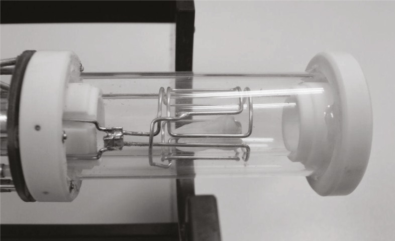
Twenty extracted human teeth were scanned on a 2.35 T MRI system for MR microscopy using the 3D spin-echo method that enabled image acquisition with isotropic resolution of 100 μm. The 3D images were then post processed by ImageJ program (NIH) to obtain advanced volume rendered views of dental pulps.
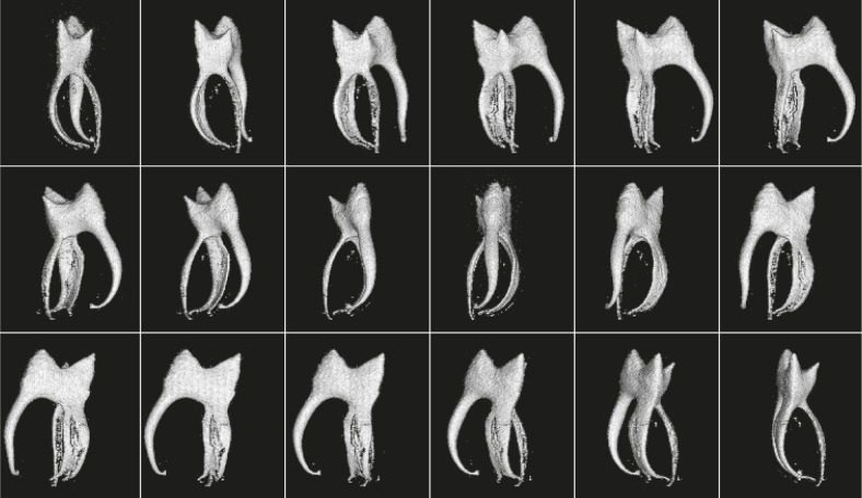
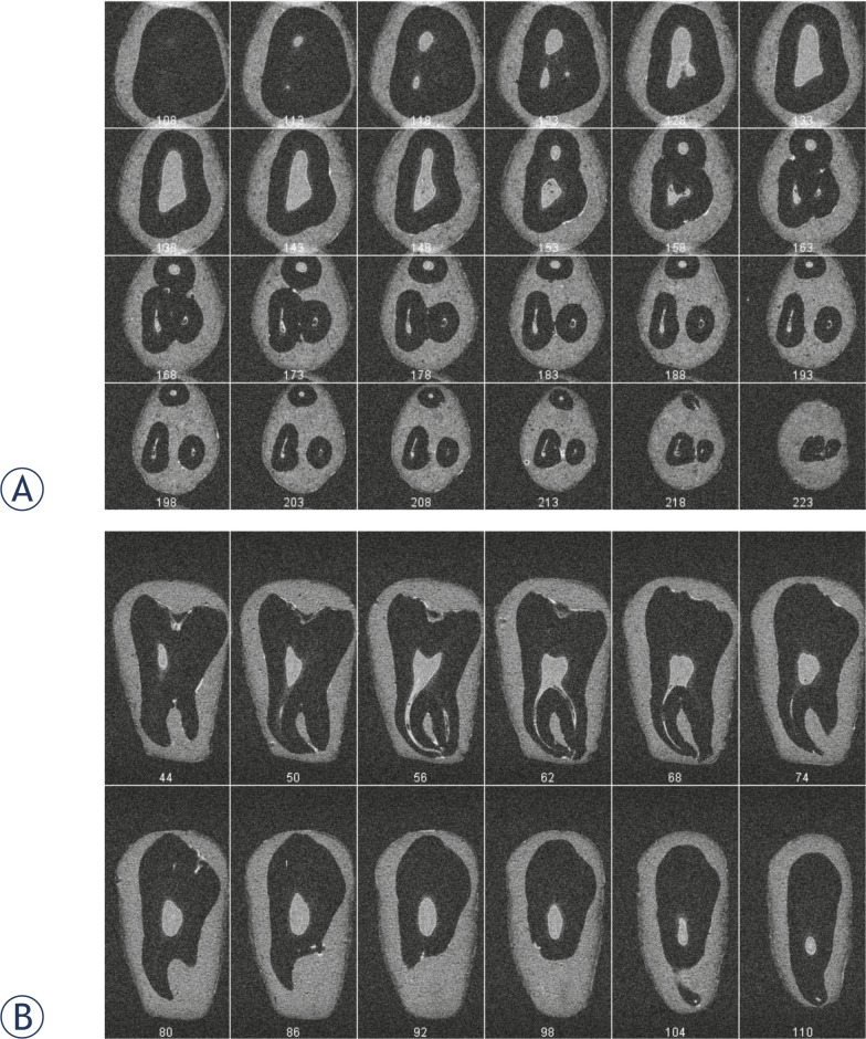
MR microscopy at 2.35 T provided accurate data on dental pulp anatomy in vitro. The data were presented as a sequence of thin 2D slices through the pulp in various orientations or as volume rendered 3D images reconstructed form arbitrary view-points. Sequential 2D images enabled only an approximate assessment of the pulp, while volume rendered 3D images were more precise in visualization of pulp anatomy and clearly showed pulp diverticles, number of pulp canals and root canal anastomosis.
This in vitro study demonstrated that MR microscopy could provide very accurate 3D visualization of dental pulp anatomy. A possible future application of the method in vivo may be of a great importance for the endodontic treatment.
精确评估牙髓解剖结构对于成功的根管治疗至关重要。由于牙齿的标准射线照相术只能提供非常有限的牙髓解剖结构信息,因此更具能力的方法备受青睐。其中之一是 3D 磁共振(MR)显微镜,该研究评估了其在评估更好的牙髓解剖结构方面的诊断能力。
使用 3D 自旋回波方法,在 2.35T MRI 系统上对 20 颗离体人牙进行扫描,以进行 MR 显微镜检查,该方法能够以 100μm 的各向同性分辨率获取图像。然后使用 ImageJ 程序(NIH)对 3D 图像进行后处理,以获得牙髓的高级容积渲染视图。
2.35T 的 MR 显微镜在体外提供了牙髓解剖结构的准确数据。这些数据以通过牙髓的各种方向的薄 2D 切片序列或从任意视角重建的容积渲染 3D 图像的形式呈现。连续的 2D 图像仅能对牙髓进行大致评估,而容积渲染的 3D 图像则更能精确地显示牙髓解剖结构,并清楚地显示出牙髓分歧、牙髓管数量和根管吻合。
这项体外研究表明,MR 显微镜可以提供非常准确的牙髓解剖 3D 可视化。该方法在体内的未来潜在应用可能对根管治疗具有重要意义。