Zhao Zhenghang, Wen Hairuo, Fefelova Nadezhda, Allen Charelle, Guillaume Nancy, Xiao Dandan, Huang Chen, Zang Weijin, Gwathmey Judith K, Xie Lai-Hua
Department of Pharmacology, School of Medicine, Xi'an Jiaotong University Xi'an China.
Front Physiol. 2012 Jul 9;3:252. doi: 10.3389/fphys.2012.00252. eCollection 2012.
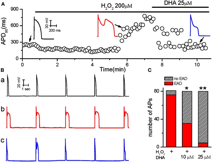
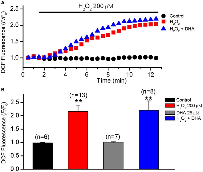
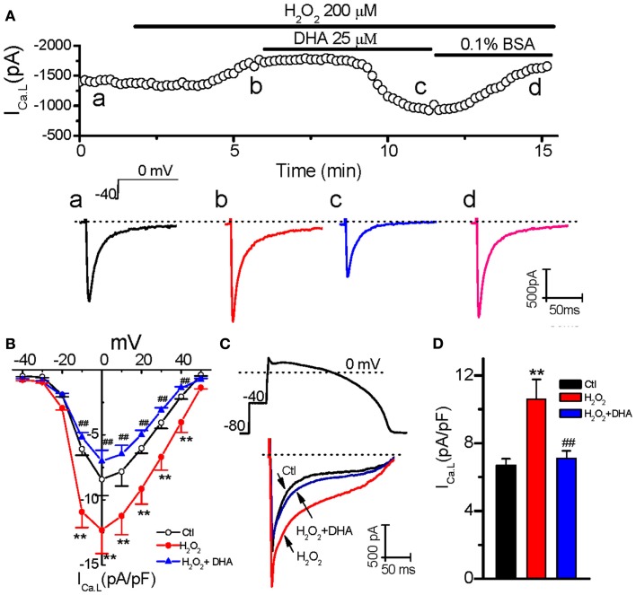
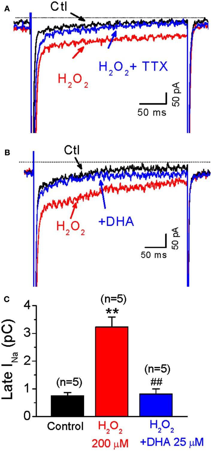
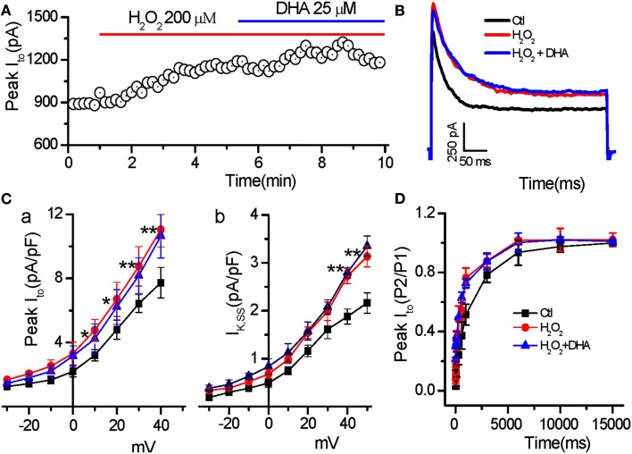
Accumulating evidence has suggested that ω3-polyunsaturated fatty acids (ω3-PUFAs) may have beneficial effects in the prevention/treatment of cardiovascular diseases, while controversies still remain regarding their anti-arrhythmic potential. It is not clear yet whether ω-3-PUFAs can suppress early afterdepolarizations (EADs) induced by oxidative stress. In the present study, we recorded action potentials using the patch-clamp technique in ventricular myocytes isolated from rabbit hearts. The treatment of myocytes with H(2)O(2) (200 μM) prolonged AP durations and induced EADs, which were significantly suppressed by docosahexaenoic acid (DHA, 10 or 25 μM; n = 8). To reveal the ionic mechanisms, we examined the effects of DHA on L-type calcium currents (I(Ca.L)), late sodium (I(Na)), and transient outward potassium currents (I(to)) in ventricular myocytes pretreated with H(2)O(2). H(2)O(2) (200 μM) increased I(Ca.L) by 46.4% from control (-8.4 ± 1.4 pA/pF) to a peak level (-12.3 ± 1.8 pA/pF, n = 6, p < 0.01) after 6 min of H(2)O(2) perfusion. H(2)O(2)-enhanced I(Ca.L) was significantly reduced by DHA (25 μM; -7.1 ± 0.9 pA/pF, n = 6, p < 0.01). Similarly, H(2)O(2)-increased the late I(Na) (-3.2 ± 0.3 pC) from control level (-0.7 ± 0.1 pC). DHA (25 μM) completely reversed the H(2)O(2)-induced increase in late I(Na) (to -0.8 ± 0.2 pC, n = 5). H(2)O(2) also increased the peak amplitude of and the steady state I(to) from 8.9 ± 1.0 and 2.16 ± 0.25 pA/pF to 12.8 ± 1.21 and 3.13 ± 0.47 pA/pF respectively (n = 6, p < 0.01, however, treatment with DHA (25 μM) did not produce significant effects on current amplitudes and dynamics of I(to) altered by H(2)O(2). In addition, DHA (25 μM) did not affect the increase of intracellular reactive oxygen species (ROS) levels induced by H(2)O(2) in rabbit ventricular myocytes. These findings demonstrate that DHA suppresses exogenous H(2)O(2)-induced EADs mainly by modulating membrane ion channel functions, while its direct effect on ROS may play a less prominent role.
越来越多的证据表明,ω3多不饱和脂肪酸(ω3-PUFAs)可能在预防/治疗心血管疾病方面具有有益作用,但其抗心律失常潜力仍存在争议。目前尚不清楚ω-3-PUFAs是否能抑制氧化应激诱导的早期后去极化(EADs)。在本研究中,我们使用膜片钳技术记录了从兔心脏分离的心室肌细胞的动作电位。用H₂O₂(200μM)处理心肌细胞可延长动作电位时程并诱导EADs,二十二碳六烯酸(DHA,10或25μM;n = 8)可显著抑制这些现象。为揭示其离子机制,我们研究了DHA对预先用H₂O₂处理的心室肌细胞L型钙电流(I(Ca.L))、晚钠电流(I(Na))和瞬时外向钾电流(I(to))的影响。H₂O₂(200μM)灌注6分钟后,I(Ca.L)从对照水平(-8.4±1.4 pA/pF)增加46.4%至峰值水平(-12.3±1.8 pA/pF,n = 6,p < 0.01)。DHA(25μM)可显著降低H₂O₂增强的I(Ca.L)(-7.1±0.9 pA/pF,n = 6,p < 0.01)。同样,H₂O₂使晚I(Na)从对照水平(-0.7±0.1 pC)增加至(-3.2±0.3 pC)。DHA(25μM)完全逆转了H₂O₂诱导的晚I(Na)增加(至-0.8±0.2 pC,n = 5)。H₂O₂还使I(to)的峰值幅度和稳态值分别从8.9±1.0和2.16±0.25 pA/pF增加至12.8±1.21和3.13±0.47 pA/pF(n = 6,p < 0.01),然而,DHA(25μM)对H₂O₂改变的I(to)的电流幅度和动力学没有显著影响。此外,DHA(25μM)不影响H₂O₂诱导的兔心室肌细胞内活性氧(ROS)水平的升高。这些发现表明,DHA主要通过调节膜离子通道功能来抑制外源性H₂O₂诱导的EADs,而其对ROS的直接作用可能不太显著。