Côté Marie-Pascale, Detloff Megan R, Wade Rodel E, Lemay Michel A, Houlé John D
Department of Neurobiology and Anatomy, Drexel University College of Medicine Philadelphia, PA, USA.
Front Physiol. 2012 Aug 17;3:330. doi: 10.3389/fphys.2012.00330. eCollection 2012.
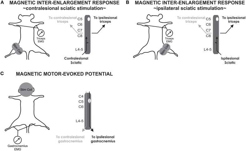
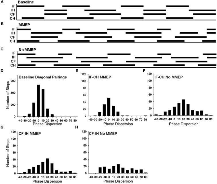
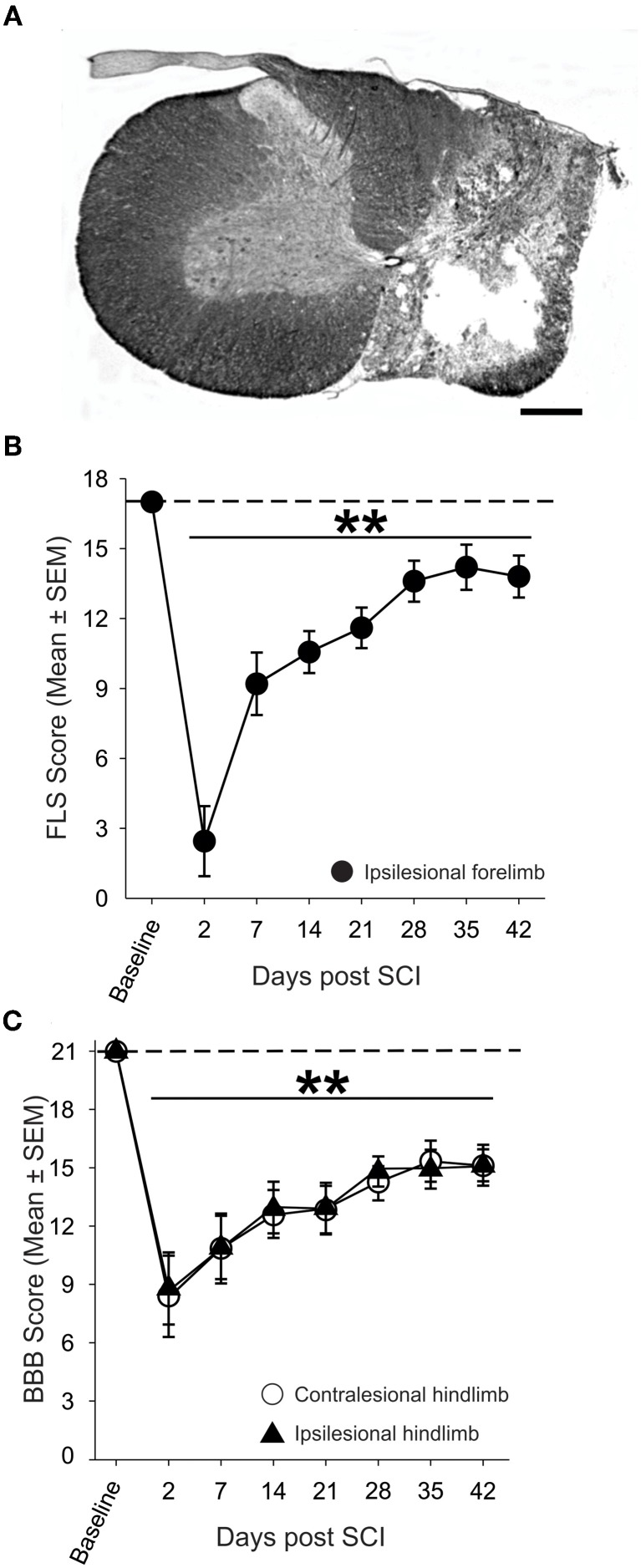
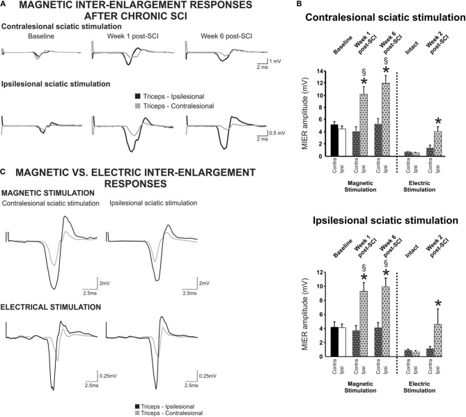
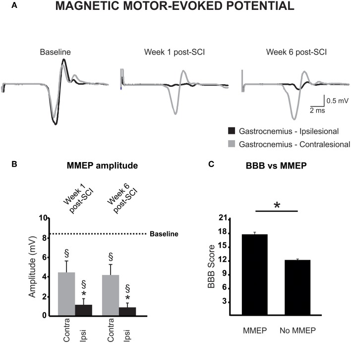
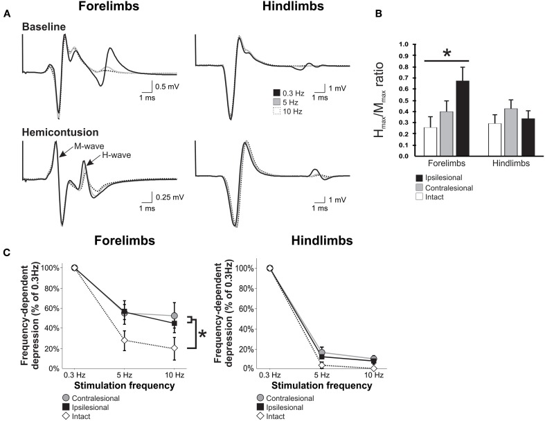
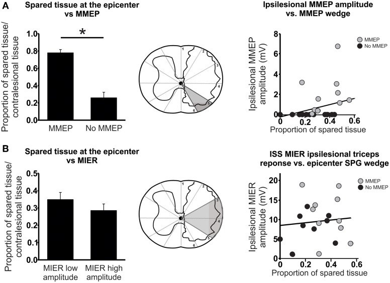
The high clinical relevance of models of incomplete cervical spinal cord injury (SCI) creates a need to address the spontaneous neuroplasticity that underlies changes in functional activity that occur over time after SCI. There is accumulating evidence supporting long projecting propriospinal neurons as suitable targets for therapeutic intervention after SCI, but focus has remained primarily oriented toward study of descending pathways. Long ascending axons from propriospinal neurons at lower thoracic and lumbar levels that form inter-enlargement pathways are involved in forelimb-hindlimb coordination during locomotion and are capable of modulating cervical motor output. We used non-invasive magnetic stimulation to assess how a unilateral cervical (C5) spinal contusion might affect transmission in intact, long ascending propriospinal pathways, and influence spinal cord plasticity. Our results show that transmission is facilitated in this pathway on the ipsilesional side as early as 1 week post-SCI. We also probed for descending magnetic motor evoked potentials (MMEPs) and found them absent or greatly reduced on the ipsilesional side as expected. The frequency-dependent depression (FDD) of the H-reflex recorded from the forelimb triceps brachii was bilaterally decreased although H(max)/M(max) was increased only on the ipsilesional side. Behaviorally, stepping recovered, but there were deficits in forelimb-hindlimb coordination as detected by BBB and CatWalk measures. Importantly, epicenter sparing correlated to the amplitude of the MMEPs and locomotor recovery but it was not significantly associated with the inter-enlargement or segmental H-reflex. In summary, our results indicate that complex plasticity occurs after a C5 hemicontusion injury, leading to differential changes in ascending vs. descending pathways, ipsi- vs. contralesional sides even though the lesion was unilateral as well as cervical vs. lumbar local spinal networks.
不完全性颈脊髓损伤(SCI)模型具有高度的临床相关性,这就需要研究SCI后随时间推移发生的功能活动变化背后的自发神经可塑性。越来越多的证据支持长投射脊髓固有神经元是SCI后治疗干预的合适靶点,但研究重点主要仍集中在下行通路。来自胸段和腰段较低水平脊髓固有神经元的长上行轴突形成节段间通路,参与运动过程中的前肢-后肢协调,并且能够调节颈段运动输出。我们使用非侵入性磁刺激来评估单侧颈(C5)脊髓挫伤如何影响完整的长上行脊髓固有通路的传导,并影响脊髓可塑性。我们的结果表明,早在SCI后1周,同侧该通路的传导就得到了促进。我们还检测了下行磁运动诱发电位(MMEP),发现如预期的那样,同侧的MMEP缺失或大幅降低。从前肢肱三头肌记录的H反射的频率依赖性抑制(FDD)在双侧均降低,尽管仅在同侧H(max)/M(max)增加。行为学上,踏步功能恢复,但通过BBB和CatWalk测量发现前肢-后肢协调存在缺陷。重要的是,损伤中心保留与MMEP的幅度和运动恢复相关,但与节段间或节段性H反射无显著关联。总之,我们的结果表明,C5半挫伤损伤后会发生复杂的可塑性变化,导致上行与下行通路、同侧与对侧、即使损伤是单侧的以及颈段与腰段局部脊髓网络之间出现不同的变化。