Department of Neuroscience, Cell Biology, and Physiology, Wright State University School of Medicine, 3640 Colonel Glenn Hwy, Dayton, OH, 45435, USA.
Skelet Muscle. 2012 Aug 30;2(1):17. doi: 10.1186/2044-5040-2-17.
During the acute phase of critical illness myopathy (CIM) there is inexcitability of skeletal muscle. In a rat model of CIM, muscle inexcitability is due to inactivation of sodium channels. A major contributor to this sodium channel inactivation is a hyperpolarized shift in the voltage dependence of sodium channel inactivation. The goal of the current study was to find a biochemical correlate of the hyperpolarized shift in sodium channel inactivation.
The rat model of CIM was generated by cutting the sciatic nerve and subsequent injections of dexamethasone for 7 days. Skeletal muscle membranes were prepared from gastrocnemius muscles, and purification and biochemical analyses carried out. Immunoprecipitations were performed with a pan-sodium channel antibody, and the resulting complexes probed in Western blots with various antibodies.
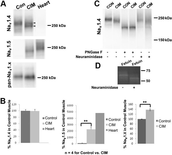
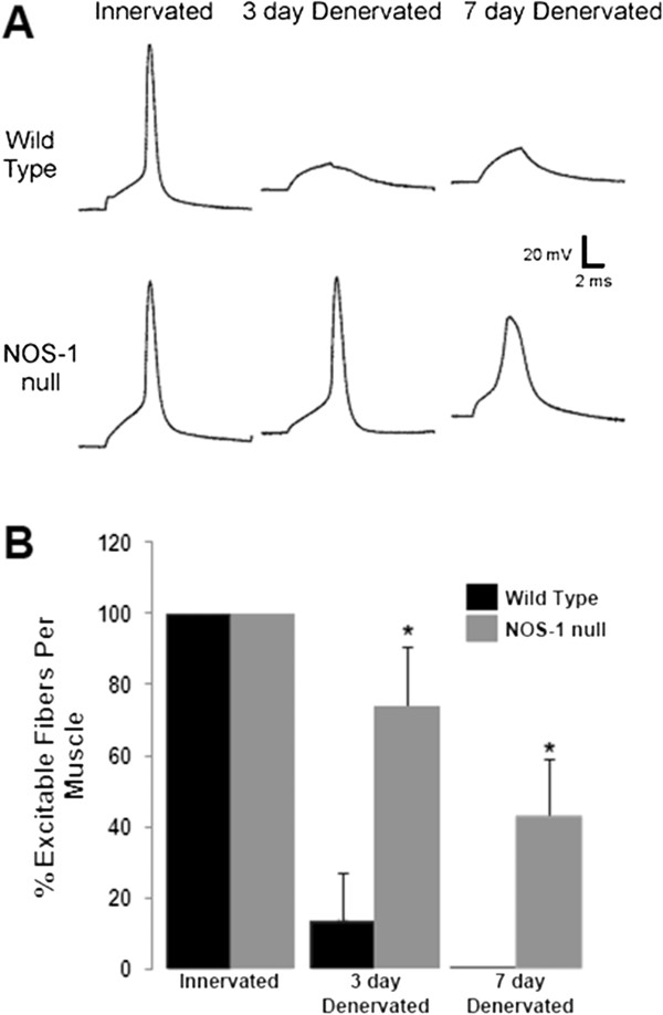
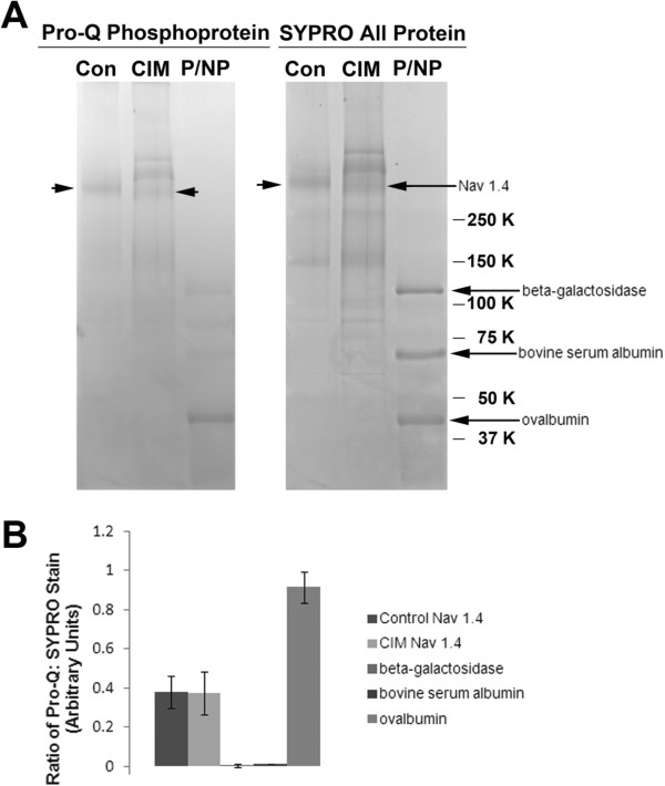
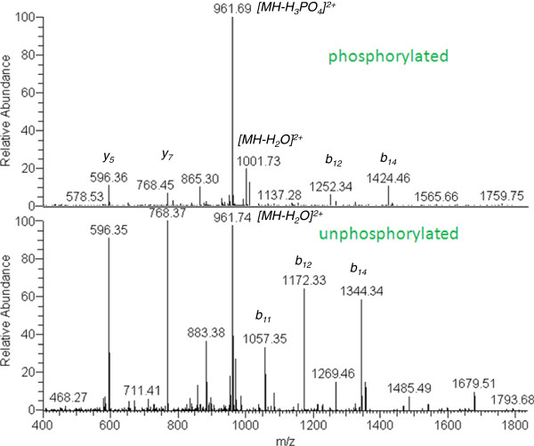
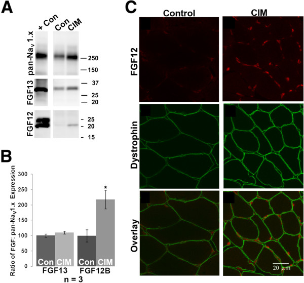
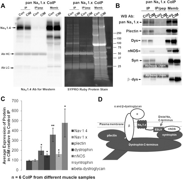
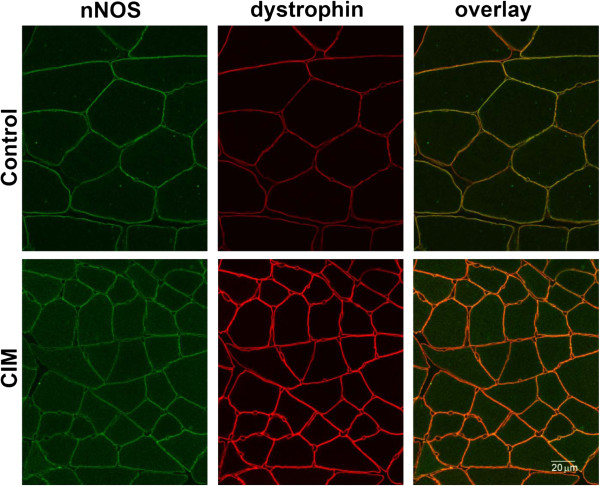
We carried out analyses of sodium channel glycosylation, phosphorylation, and association with other proteins. Although there was some loss of channel glycosylation in the disease, as assessed by size analysis of glycosylated and de-glycosylated protein in control and CIM samples, previous work by other investigators suggest that such loss would most likely shift channel inactivation gating in a depolarizing direction; thus such loss was viewed as compensatory rather than causative of the disease. A phosphorylation site at serine 487 was identified on the NaV 1.4 sodium channel α subunit, but there was no clear evidence of altered phosphorylation in the disease. Co-immunoprecipitation experiments carried out with a pan-sodium channel antibody confirmed that the sodium channel was associated with proteins of the dystrophin associated protein complex (DAPC). This complex differed between control and CIM samples. Syntrophin, dystrophin, and plectin associated strongly with sodium channels in both control and disease conditions, while β-dystroglycan and neuronal nitric oxide synthase (nNOS) associated strongly with the sodium channel only in CIM. Recording of action potentials revealed that denervated muscle in mice lacking nNOS was more excitable than control denervated muscle.
Taken together, these data suggest that the conformation/protein association of the sodium channel complex differs in control and critical illness myopathy muscle membranes; and suggest that nitric oxide signaling plays a role in development of muscle inexcitability.
在危重病性肌病(CIM)的急性期,骨骼肌无兴奋性。在 CIM 的大鼠模型中,肌肉无兴奋性是由于钠通道失活。导致这种钠通道失活的主要原因是钠通道失活的电压依赖性发生超极化漂移。本研究的目的是寻找钠通道失活超极化漂移的生化相关物。
通过切断坐骨神经和随后注射地塞米松 7 天来建立 CIM 的大鼠模型。从比目鱼肌中制备骨骼肌膜,并进行纯化和生化分析。用泛钠通道抗体进行免疫沉淀,然后用各种抗体在 Western 印迹中探测所得复合物。
我们进行了钠通道糖基化、磷酸化和与其他蛋白相互作用的分析。尽管在疾病中观察到通道糖基化的一些损失,如通过控制和 CIM 样本中糖基化和去糖基化蛋白的大小分析来评估,但其他研究人员的先前工作表明,这种损失很可能使通道失活门控向去极化方向移动;因此,这种损失被视为代偿性的,而不是疾病的原因。在 NaV1.4 钠通道α亚基上鉴定出丝氨酸 487 位的磷酸化位点,但在疾病中没有明显的磷酸化证据。用泛钠通道抗体进行的共免疫沉淀实验证实,钠通道与营养不良相关蛋白复合物(DAPC)的蛋白相关。该复合物在对照和疾病样本之间存在差异。在对照和疾病条件下,联蛋白、营养不良蛋白和网蛋白与钠通道强烈结合,而β-肌营养不良蛋白和神经元型一氧化氮合酶(nNOS)仅在 CIM 中与钠通道强烈结合。动作电位记录显示,缺乏 nNOS 的去神经肌肉比对照去神经肌肉更兴奋。
综上所述,这些数据表明,钠通道复合物的构象/蛋白相互作用在对照和危重病性肌病肌肉膜中存在差异;并表明一氧化氮信号在肌肉无兴奋性的发展中起作用。