Department of Radiology, New York University School of Medicine, 660 First Avenue, New York, NY 10016, USA.
Cancer Imaging. 2012 Aug 10;12(1):237-44. doi: 10.1102/1470-7330.2012.0028.
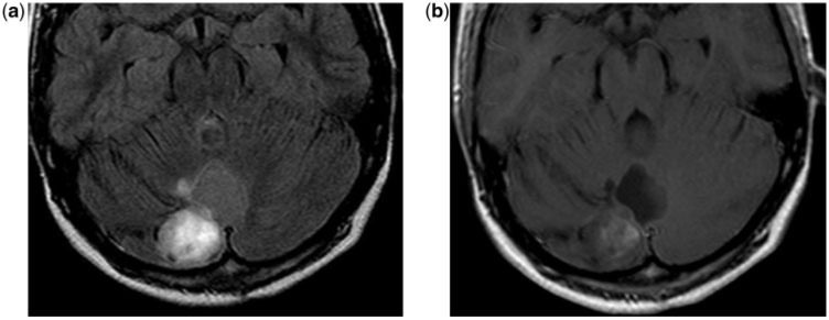
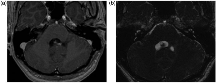
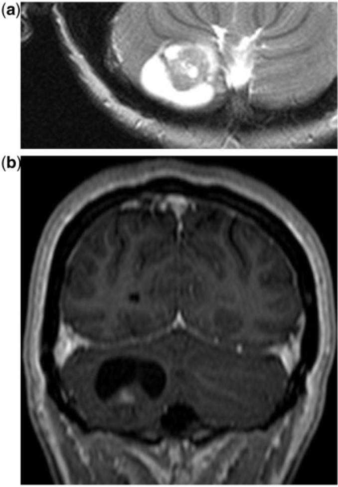
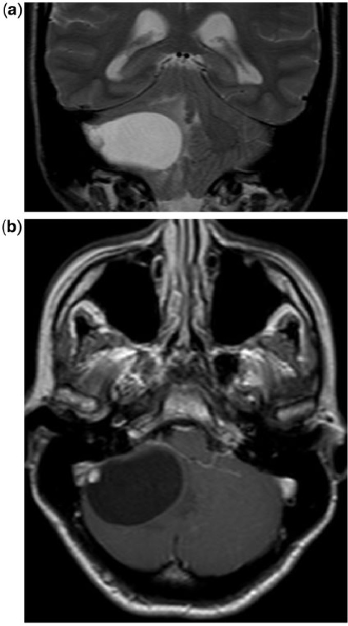
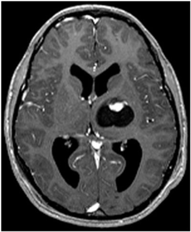
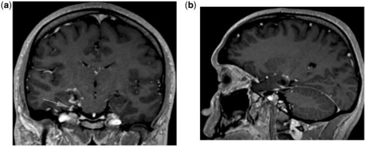
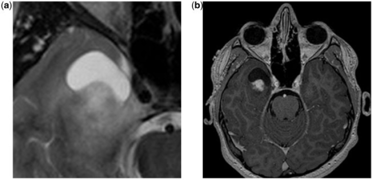
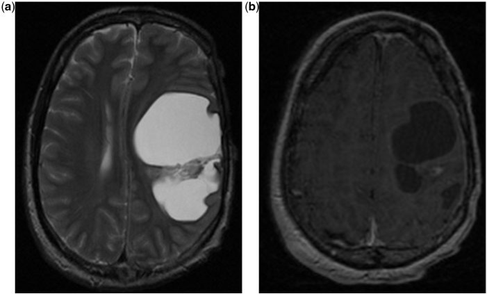
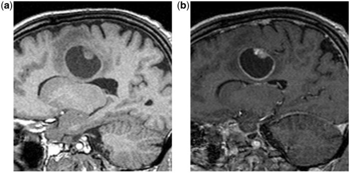
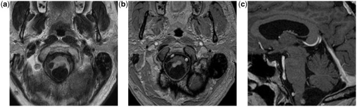
The purpose of this article is to illustrate the imaging findings of lesions that present as cyst with a mural nodule tumor (CMNT). CMNT is a subtype pattern of intra-axial enhancement in central nervous system tumors, typical of a variety of brain neoplasms, including, as the most common, hemangioblastoma, pilocytic astrocytoma, ganglioglioma and pleomorphic xanthoastrocytoma and as less common tanycytic ependymoma, intraparenchymal schwannoma, desmoplastic infantile ganglioglioma and cystic metastasis. A retrospective design was chosen given the rarity of CMNT. Relevant cases were obtained retrospectively to review the different lesions that can present with the appearance of CMNT.
本文旨在阐述以囊壁结节肿瘤(CMNT)为表现的病变的影像学特征。CMNT 是中枢神经系统肿瘤的一种轴内强化亚型模式,典型表现为多种脑肿瘤,包括最常见的血管母细胞瘤、毛细胞型星形细胞瘤、神经节胶质瘤和多形性黄色星形细胞瘤,以及较少见的室管膜下瘤、脑实质内 Schwann 细胞瘤、促结缔组织增生性婴儿型神经节胶质瘤和囊性转移瘤。鉴于 CMNT 的罕见性,选择了回顾性设计。回顾性获得了相关病例,以探讨可能表现为 CMNT 外观的不同病变。