Yoda Rebecca A, Cimino Patrick J
Department of Laboratory Medicine and Pathology, Division of Neuropathology, University of Washington, Seattle, WA, USA.
Department of Laboratory Medicine and Pathology, Division of Cytopathology, University of Washington, Seattle, WA, USA.
J Pathol Transl Med. 2022 May;56(3):115-125. doi: 10.4132/jptm.2022.04.13. Epub 2022 May 3.
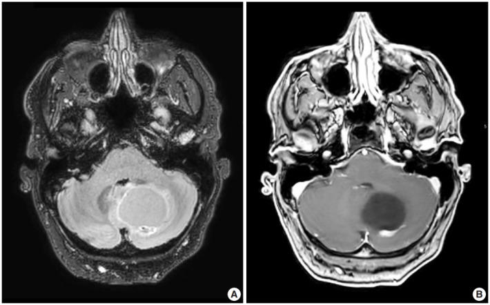
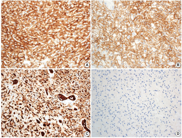
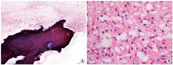
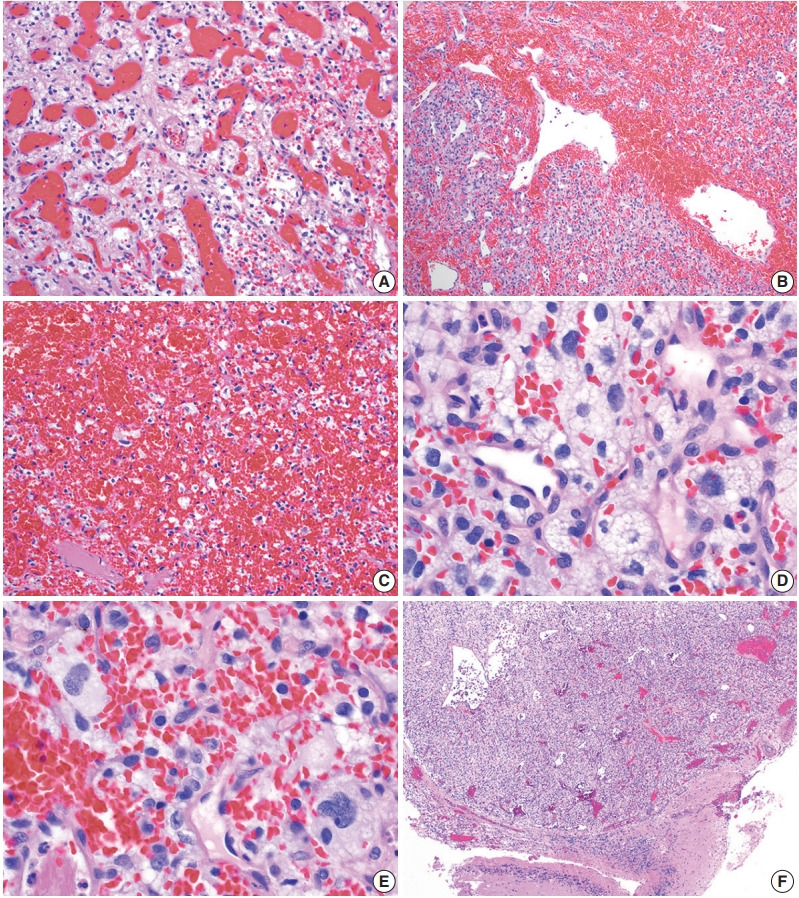
Hemangioblastoma is a benign, highly vascularized neoplasm of the central nervous system (CNS). This tumor is associated with loss of function of the VHL gene and demonstrates frequent occurrence in von Hippel-Lindau (VHL) disease. While this entity is designated CNS World Health Organization grade 1, due to its predilection for the cerebellum, brainstem, and spinal cord, it is still an important cause of morbidity and mortality in affected patients. Recognition and accurate diagnosis of hemangioblastoma is essential for the practice of surgical neuropathology. Other CNS neoplasms, including several tumors associated with VHL disease, may present as histologic mimics, making diagnosis challenging. We outline key clinical and radiologic features, pathophysiology, treatment modalities, and prognostic information for hemangioblastoma, and provide a thorough review of the gross, microscopic, immunophenotypic, and molecular features used to guide diagnosis.
血管母细胞瘤是中枢神经系统(CNS)的一种良性、高度血管化的肿瘤。该肿瘤与VHL基因功能丧失有关,且在冯·希佩尔-林道(VHL)病中经常出现。虽然该实体被世界卫生组织指定为CNS 1级,但由于其好发于小脑、脑干和脊髓,它仍是受影响患者发病和死亡的重要原因。血管母细胞瘤的识别和准确诊断对于神经外科病理学实践至关重要。其他CNS肿瘤,包括几种与VHL病相关的肿瘤,可能表现为组织学上的模仿,这使得诊断具有挑战性。我们概述了血管母细胞瘤的关键临床和放射学特征、病理生理学、治疗方式和预后信息,并对用于指导诊断的大体、显微镜、免疫表型和分子特征进行了全面综述。