Department of Physiology, Institute of Cardiovascular Sciences, St Boniface Hospital Research, Winnipeg, Manitoba, Canada.
J Cell Mol Med. 2012 Dec;16(12):2958-67. doi: 10.1111/j.1582-4934.2012.01623.x.
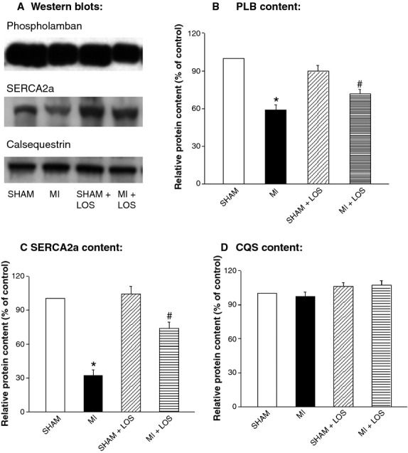
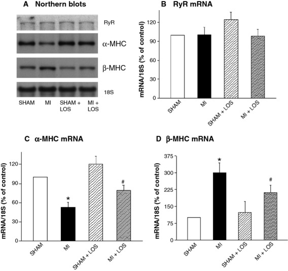
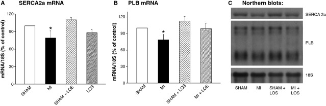
This study tested the reversal of subcellular remodelling in heart failure due to myocardial infarction (MI) upon treatment with losartan, an angiotensin II receptor antagonist. Twelve weeks after inducing MI, rats were treated with or without losartan (20 mg/kg; daily) for 8 weeks and assessed for cardiac function, cardiac remodelling, subcellular alterations and plasma catecholamines. Cardiac hypertrophy and lung congestion in 20 weeks MI-induced heart failure were associated with increases in plasma catecholamine levels. Haemodynamic examination revealed depressed cardiac function, whereas echocardiographic analysis showed impaired cardiac performance and marked increases in left ventricle wall thickness and chamber dilatation at 20 weeks of inducing MI. These changes in cardiac function, cardiac remodelling and plasma dopamine levels in heart failure were partially or fully reversed by losartan. Sarcoplasmic reticular (SR) Ca(2+)-pump activity and protein expression, protein and gene expression for phospholamban, as well as myofibrillar (MF) Ca(2+)-stimulated ATPase activity and α-myosin heavy chain mRNA levels were depressed, whereas β-myosin heavy chain expression was increased in failing hearts; these alterations were partially reversed by losartan. Although SR Ca(2+)-release activity and mRNA levels for SR Ca(2+)-pump were decreased in failing heart, these changes were not reversed upon losartan treatment; no changes in mRNA levels for SR Ca(2+)-release channels were observed in untreated or treated heart failure. These results suggest that the partial improvement of cardiac performance in heart failure due to MI by losartan treatment is associated with partial reversal of cardiac remodelling as well as partial recovery of SR and MF functions.
本研究旨在探讨血管紧张素 II 受体拮抗剂氯沙坦逆转心肌梗死后心力衰竭引起的亚细胞重构的作用。心肌梗死后 12 周,给予大鼠氯沙坦(20mg/kg;每日)治疗 8 周,评估心功能、心脏重构、亚细胞改变和血浆儿茶酚胺水平。20 周心肌梗死后心力衰竭大鼠的心脏肥大和肺淤血与血浆儿茶酚胺水平升高相关。血流动力学检查显示心功能降低,而超声心动图分析显示心功能受损,左心室壁厚度和腔室扩张明显增加。氯沙坦部分或完全逆转了心力衰竭时心脏功能、心脏重构和血浆多巴胺水平的改变。肌浆网(SR)Ca2+-泵活性和蛋白表达、磷蛋白表达、肌球蛋白轻链(MF)Ca2+刺激 ATP 酶活性和α-肌球蛋白重链 mRNA 水平降低,而β-肌球蛋白重链表达增加;氯沙坦部分逆转了这些改变。尽管 SR Ca2+-释放活性和 SR Ca2+-泵的 mRNA 水平在心力衰竭时降低,但氯沙坦治疗并未逆转这些变化;未观察到 SR Ca2+释放通道的 mRNA 水平在未治疗或治疗的心力衰竭中发生变化。这些结果表明,氯沙坦治疗部分改善心肌梗死后心力衰竭的心脏功能与心脏重构的部分逆转以及 SR 和 MF 功能的部分恢复有关。