Faculdade de Medicina, Universidade de São Paulo, São Paulo, SP, Brazil.
Clinics (Sao Paulo). 2012 Aug;67(8):931-7. doi: 10.6061/clinics/2012(08)13.
Acute retinal necrosis is a rapidly progressive and devastating viral retinitis caused by the herpesvirus family. Systemic acyclovir is the treatment of choice; however, the progression of retinal lesions ceases approximately 2 days after treatment initiation. An intravitreal injection of acyclovir may be used an adjuvant therapy during the first 2 days of treatment when systemically administered acyclovir has not reached therapeutic levels in the retina. The aims of this study were to determine the pharmacokinetic profile of acyclovir in the rabbit vitreous after intravitreal injection and the functional effects of acyclovir in the rabbit retina.
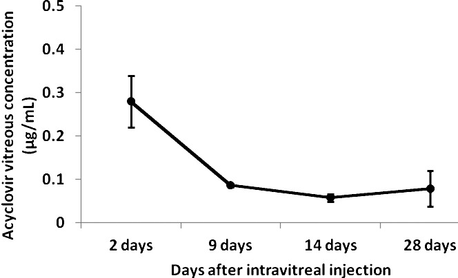
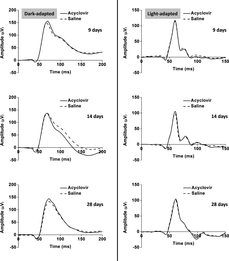
Acyclovir (Acyclovir; Bedford Laboratories, Bedford, OH, USA) 1 mg in 0.1 mL was injected into the right eye vitreous of 32 New Zealand white rabbits, and 0.1 mL sterile saline solution was injected into the left eye as a control. The animals were sacrificed after 2, 9, 14, or 28 days. The eyes were enucleated, and the vitreous was removed. The half-life of acyclovir was determined using high-performance liquid chromatography. Electroretinograms were recorded on days 2, 9, 14, and 28 in the eight animals that were sacrificed 28 days after injection according to a modified protocol of the International Society for Clinical Electrophysiology of Vision.
Acyclovir rapidly decayed in the vitreous within the first two days after treatment and remained at low levels from day 9 onward. The eyes that were injected with acyclovir did not present any electroretinographic changes compared with the control eyes.
The vitreous half-life of acyclovir is short, and the electrophysiological findings suggest that the intravitreal delivery of 1 mg acyclovir is safe and well tolerated by the rabbit retina.
急性视网膜坏死是一种由疱疹病毒家族引起的快速进展和破坏性的病毒性视网膜炎。系统使用阿昔洛韦是治疗的首选;然而,视网膜病变的进展在开始治疗后约 2 天停止。当全身性给予阿昔洛韦在视网膜中未达到治疗水平时,在治疗的头 2 天内,可以使用玻璃体内注射阿昔洛韦作为辅助治疗。本研究的目的是确定玻璃体内注射阿昔洛韦后兔玻璃体内的药代动力学特征,以及阿昔洛韦对兔视网膜的功能影响。
将 1 毫克阿昔洛韦(阿昔洛韦;贝德福德实验室,贝德福德,俄亥俄州,美国)溶于 0.1 毫升中,注入 32 只新西兰白兔的右眼玻璃体,左眼注射 0.1 毫升无菌生理盐水作为对照。在第 2、9、14 或 28 天处死动物。眼球被切除,取出玻璃体。使用高效液相色谱法确定阿昔洛韦的半衰期。根据国际临床视觉电生理学协会的改良方案,在注射后 28 天处死的 8 只动物中,于第 2、9、14 和 28 天记录视网膜电图。
阿昔洛韦在治疗后的头两天内迅速在玻璃体内降解,并且从第 9 天开始一直保持低水平。与对照眼相比,注射阿昔洛韦的眼睛没有任何视网膜电图变化。
阿昔洛韦的玻璃体半衰期短,并且电生理发现表明,玻璃体内给予 1 毫克阿昔洛韦是安全的,兔视网膜可以很好地耐受。