Department of Cancer Biology, The University of Texas MD Anderson Cancer Center, Houston, Texas, USA.
PLoS One. 2012;7(8):e44033. doi: 10.1371/journal.pone.0044033. Epub 2012 Aug 27.
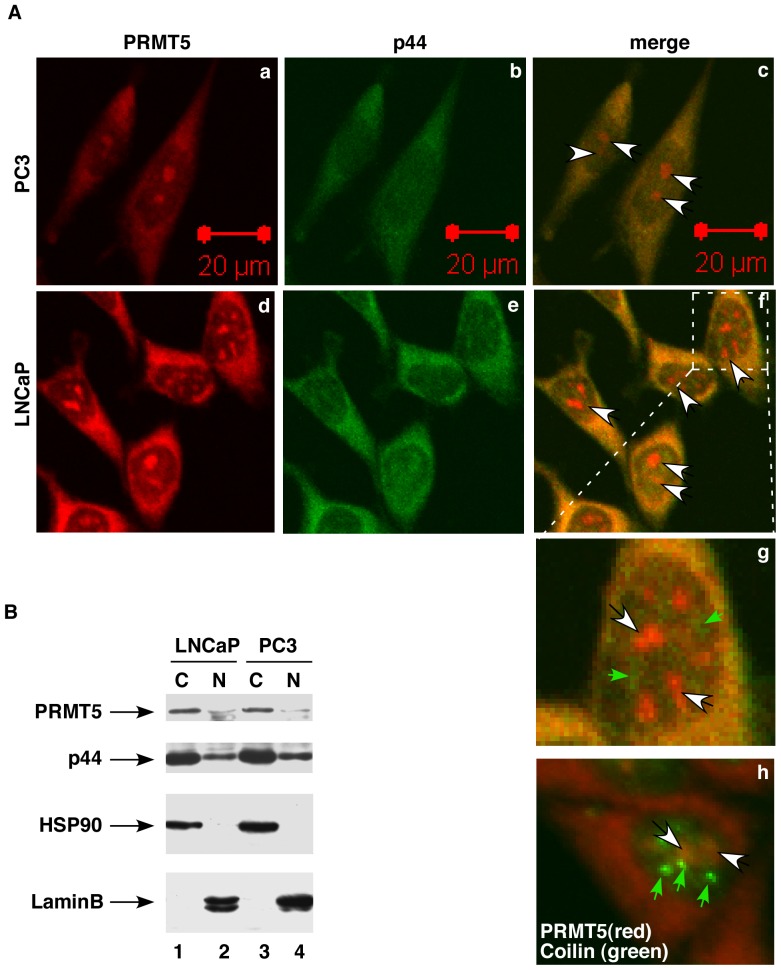
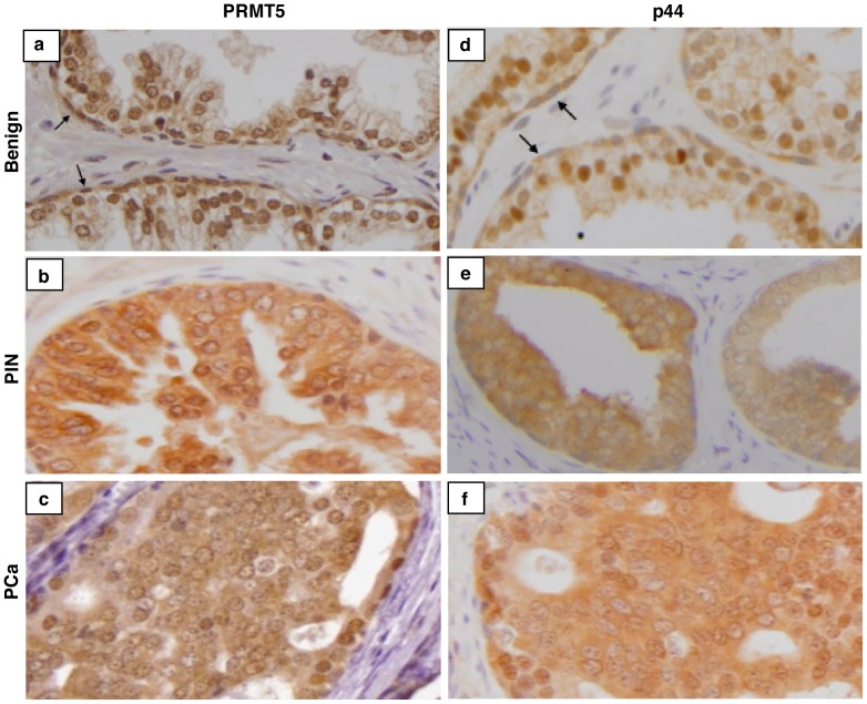
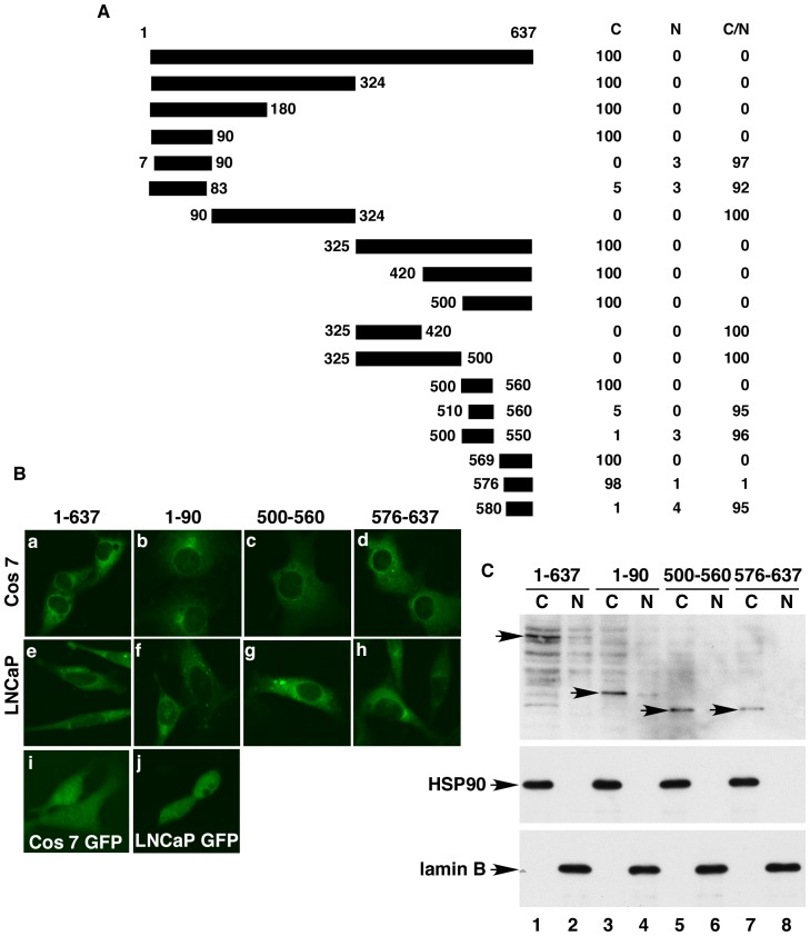
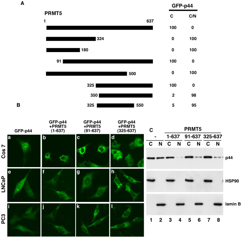
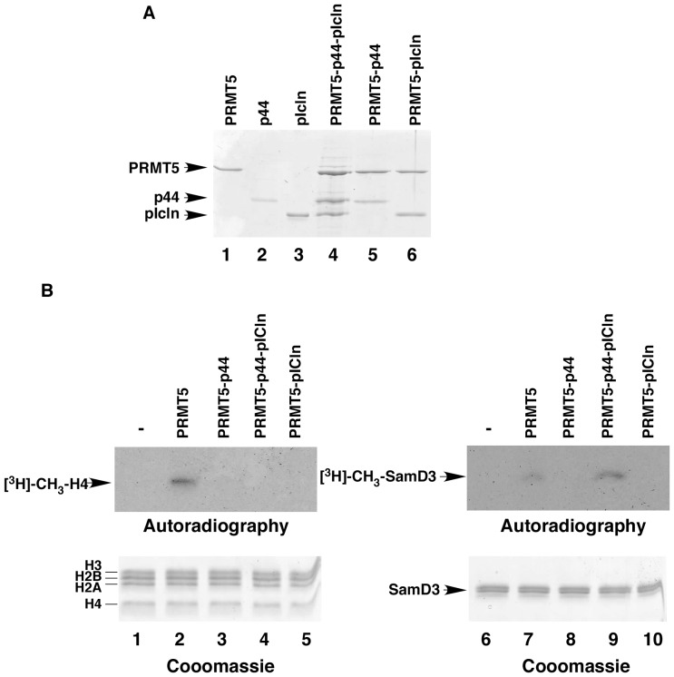
Protein arginine methyltransferase 5 (PRMT5) plays multiple roles in a large number of cellular processes, and its subcellular localization is dynamically regulated during mouse development and cellular differentiation. However, little is known of the functional differences between PRMT5 in the cytoplasm and PRMT5 in the nucleus. Here, we demonstrated that PRMT5 predominantly localized in the cytoplasm of prostate cancer cells. Subcellular localization assays designed to span the entire open-reading frame of the PRMT5 protein revealed the presence of three nuclear exclusion signals (NESs) in the PRMT5 protein. PRMT5 and p44/MED50/WD45/WDR77 co-localize in the cytoplasm, and both are required for the growth of prostate cancer cells in an PRMT5 methyltransferase activity-dependent manner. In contrast, PRMT5 in the nucleus inhibited cell growth in a methyltransferase activity-independent manner. Consistent with these observations, PRMT5 localized in the nucleus in benign prostate epithelium, whereas it localized in the cytoplasm in prostate premalignant and cancer tissues. We further found that PRMT5 alone methylated both histone H4 and SmD3 proteins but PRMT5 complexed with p44 and pICln methylated SmD3 but not histone H4. These results imply a novel mechanism by which PRMT5 controls cell growth and contributes to prostate tumorigenesis.
蛋白质精氨酸甲基转移酶 5(PRMT5)在大量细胞过程中发挥多种作用,其亚细胞定位在小鼠发育和细胞分化过程中是动态调节的。然而,对于细胞质中的 PRMT5 和核中的 PRMT5 之间的功能差异知之甚少。在这里,我们证明 PRMT5 主要定位于前列腺癌细胞的细胞质中。设计跨越 PRMT5 蛋白全长的亚细胞定位测定法揭示了 PRMT5 蛋白中存在三个核排斥信号(NES)。PRMT5 和 p44/MED50/WD45/WDR77 在细胞质中共定位,并且都需要 PRMT5 甲基转移酶活性依赖性方式来促进前列腺癌细胞的生长。相比之下,核中的 PRMT5 以甲基转移酶活性非依赖性方式抑制细胞生长。与这些观察结果一致,PRMT5 在良性前列腺上皮中定位于核内,而在前列腺前恶性和癌症组织中定位于细胞质中。我们进一步发现 PRMT5 单独甲基化组蛋白 H4 和 SmD3 蛋白,但 PRMT5 与 p44 和 pICln 复合甲基化 SmD3 但不甲基化组蛋白 H4。这些结果暗示了 PRMT5 控制细胞生长并促进前列腺肿瘤发生的新机制。