Kim Ki-Song, Byun Min-Kwang, Lee Won-Hwee, Cynn Heon-Seock, Kwon Oh-Yun, Yi Chung-Hwi
Department of Physical Therapy, The Graduate School, Yonsei University, 1 Yonseidae-gil, Wonju, Gangwon-do, 220-710, South Korea.
Division of Pulmonary Medicine, Department of Internal Medicine, Gangnam Severance Hospital, Yonsei University College of Medicine, Yonsei University Health System, 211 Eonju-ro, Gangnam-gu, Seoul 135-720, South Korea.
Multidiscip Respir Med. 2012 Jun 20;7(1):9. doi: 10.1186/2049-6958-7-9.
To determine the influence of breathing maneuver and sitting posture on tidal volume (TV), respiratory rate (RR), and muscle activity of the inspiratory accessory muscles in patients with chronic obstructive pulmonary disease (COPD).
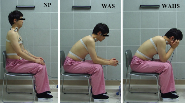
Twelve men with COPD participated in the study. Inductive respiratory plethysmography and surface electromyography were used to simultaneously measure TV, RR, and muscle activity of the inspiratory accessory muscles [the scalenus (SM), sternocleidomastoid (SCM), and pectoralis major (PM) muscles] during quiet natural breathing (QB) and pursed-lips breathing (PLB) in three sitting postures: neutral position (NP), with armm support (WAS), and with arm and head support (WAHS).
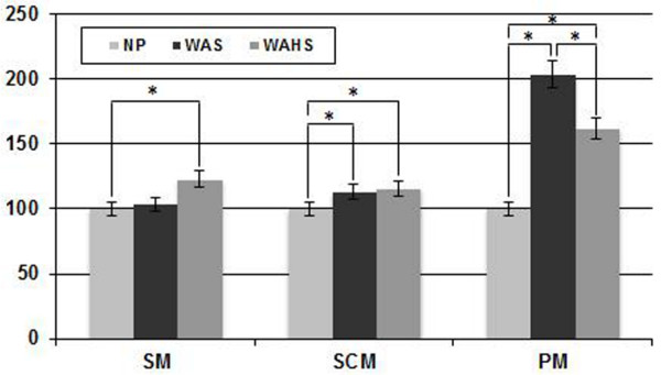
Two-way repeated-measures analysis of variance was employed. In a comparison of breathing patterns, PLB significantly increased TV and decreased RR compared to QB. Muscle activity in the SM and SCM increased significantly in PLB compared to QB. In a comparison of sitting postures, the muscle activity of the SM, SCM, and PM increased in the forward-leaning position.
The results suggest that in COPD, PLB induced a favorable breathing pattern (increased TV and reduced RR) compared to QB. Additionally, WAS and WAHS positions increased muscle activity of the inspiratory accessory muscles during inspiration versus NP. Differential involvement of accessory respiratory muscles can be readily studied in COPD patients, allowing monitoring of respiratory load during pulmonary rehabilitation.
确定呼吸动作和坐姿对慢性阻塞性肺疾病(COPD)患者潮气量(TV)、呼吸频率(RR)及吸气辅助肌肌肉活动的影响。
12名COPD男性患者参与了本研究。在三种坐姿(中立位(NP)、有手臂支撑(WAS)和有手臂及头部支撑(WAHS))下,采用感应式呼吸体积描记法和表面肌电图,在安静自然呼吸(QB)和缩唇呼吸(PLB)过程中同时测量TV、RR及吸气辅助肌(斜角肌(SM)、胸锁乳突肌(SCM)和胸大肌(PM))的肌肉活动。
采用双向重复测量方差分析。在呼吸模式比较中,与QB相比,PLB显著增加TV并降低RR。与QB相比,PLB时SM和SCM的肌肉活动显著增加。在坐姿比较中,前倾位时SM、SCM和PM的肌肉活动增加。
结果表明,在COPD患者中,与QB相比,PLB诱导出更有利的呼吸模式(TV增加和RR降低)。此外,与NP相比,WAS和WAHS位在吸气时增加了吸气辅助肌的肌肉活动。在COPD患者中可方便地研究呼吸辅助肌的不同参与情况,从而在肺康复期间监测呼吸负荷。