Department of Ophthalmology, Keio University School of Medicine, Tokyo, Japan.
PLoS One. 2012;7(9):e43688. doi: 10.1371/journal.pone.0043688. Epub 2012 Sep 4.
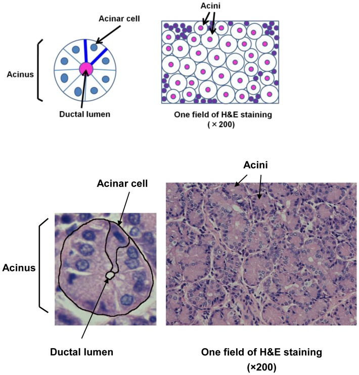
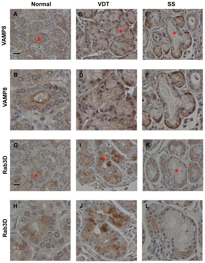
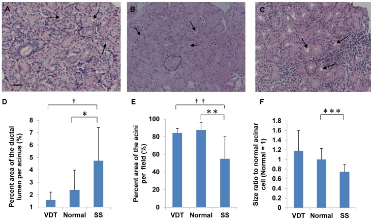
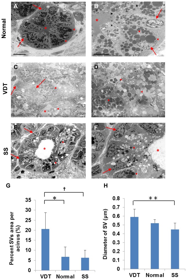
Previous observations in a rat model of a non-Sjögren's syndrome (non-SS) type of dry eye seen in users of visual display terminals (VDT) indicated that secretory vesicle (SV) accumulation in the lacrimal gland epithelia contributes to the condition. Here, to examine this possibility in humans, we compared the lacrimal gland histology and percent SV area in the cytoplasm of acinar epithelial cells using light microscopy and transmission electron microscopy, in patients with VDT work-related non-SS dry-eye (VDT group), SS-induced dry-eye, and autopsied normal controls. In addition, the VAMP8 (vesicle-associated membrane protein 8, an exocrine-pathway molecule) and Rab3D (mature vesicle marker) were histochemically examined in lacrimal gland tissue sections. The lacrimal gland acini were larger in the VDT group than in the SS group, and the percent SV area was significantly higher in the VDT group than in the normal controls (P = 0.021) or SS group (P = 0.004). Immunostaining revealed abnormal distributions of VAMP8 in the VDT and SS groups. Rab3D was more strongly expressed in the cytoplasm of acinar epithelial cells in the VDT group than in that of normal controls. The duration of VDT use was significantly longer in the VDT group than in the other groups. These findings suggest that excessive SV accumulation in the acinar epithelia may contribute to the reduced tear secretion in VDT users.
先前在使用视觉显示终端 (VDT) 的非干燥综合征 (非 SS) 型干眼症大鼠模型中的观察表明,泪腺上皮中的分泌囊泡 (SV) 积累导致了这种情况。在这里,为了在人类中检验这种可能性,我们通过光镜和透射电镜比较了 VDT 与工作相关的非 SS 干眼症 (VDT 组)、SS 诱导的干眼症和尸检正常对照组患者的泪腺组织学和空泡细胞质中 SV 区域的百分比。此外,还在泪腺组织切片中对 VAMP8(囊泡相关膜蛋白 8,外分泌途径分子)和 Rab3D(成熟囊泡标记物)进行了组织化学检查。VDT 组的泪腺腺泡比 SS 组大,SV 区域的百分比在 VDT 组明显高于正常对照组 (P = 0.021) 或 SS 组 (P = 0.004)。免疫染色显示 VDT 和 SS 组中 VAMP8 的分布异常。Rab3D 在 VDT 组的腺泡上皮细胞的细胞质中表达比正常对照组更强。VDT 使用时间在 VDT 组明显长于其他组。这些发现表明,腺泡上皮中 SV 的过度积累可能导致 VDT 用户的泪液分泌减少。