Internal Medicine, Texas Tech University Health Sciences Center, Amarillo, TX 79106, USA.
BMC Nephrol. 2012 Sep 11;13:109. doi: 10.1186/1471-2369-13-109.
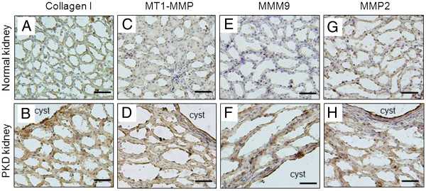
Polycystic Kidney Disease (PKD) kidneys exhibit increased extracellular matrix (ECM) collagen expression and metalloproteinases (MMPs) activity. We investigated the role of these increases on cystic disease progression in PKD kidneys.
We examined the role of type I collagen (collagen I) and membrane bound type 1 MMP (MT1-MMP) on cyst development using both in vitro 3 dimensional (3D) collagen gel culture and in vivo PCK rat model of PKD.
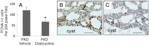
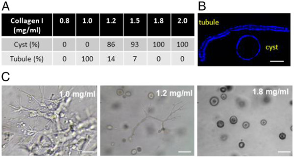
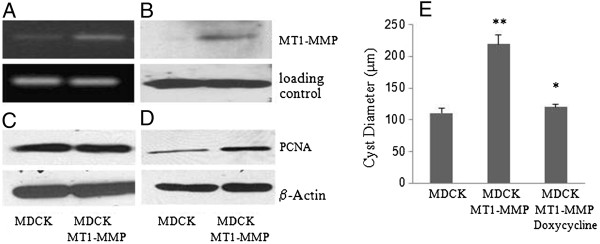
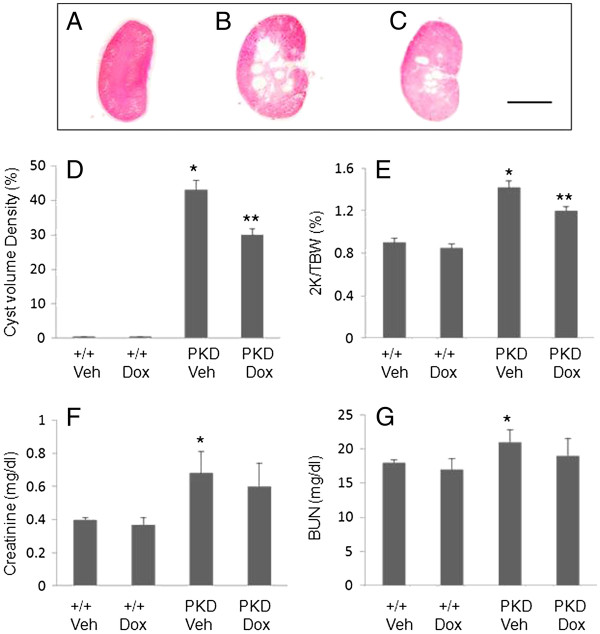
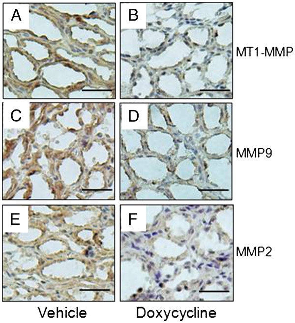
We found that collagen concentration is critical in controlling the morphogenesis of MDCK cells cultured in 3D gels. MDCK cells did not form 3D structures at collagen I concentrations lower than 1 mg/ml but began forming tubules when the concentration reaches 1 mg/ml. Significantly, these cells began to form cyst when collagen I concentration reached to 1.2 mg/ml, and the ratios of cyst to tubule structures increased as the collagen I concentration increased. These cells exclusively formed cyst structures at a collagen I concentration of 1.8 mg/ml or higher. Overexpression of MT1-MMP in MDCK cells significantly induced cyst growth in 3D collagen gel culture. Conversely, inhibition of MMPs activity with doxycycline, a FDA approved pan-MMPs inhibitor, dramatically slowed cyst growth. More importantly, the treatment of PCK rats with doxycycline significantly decreased renal tubule cell proliferation and markedly inhibited the cystic disease progression.
Our data suggest that increased collagen expression and MMP activity in PKD kidneys may induce cyst formation and expansion. Our findings also suggest that MMPs may serve as a therapeutic target for the treatment of human PKD.
多囊肾病(PKD)肾脏表现出细胞外基质(ECM)胶原表达增加和基质金属蛋白酶(MMPs)活性增强。我们研究了这些增加在 PKD 肾脏囊性疾病进展中的作用。
我们使用体外 3 维(3D)胶原凝胶培养和体内 PCK 大鼠 PKD 模型,研究了 I 型胶原(胶原 I)和膜结合型 1 MMP(MT1-MMP)对囊肿形成的作用。
我们发现胶原浓度在控制 MDCK 细胞在 3D 凝胶中培养的形态发生中起关键作用。当胶原 I 浓度低于 1mg/ml 时,MDCK 细胞不会形成 3D 结构,但当浓度达到 1mg/ml 时,细胞开始形成小管。重要的是,当胶原 I 浓度达到 1.2mg/ml 时,这些细胞开始形成囊肿,并且随着胶原 I 浓度的增加,囊肿与小管结构的比例增加。当胶原 I 浓度达到 1.8mg/ml 或更高时,这些细胞仅形成囊肿结构。MT1-MMP 在 MDCK 细胞中的过表达显著诱导 3D 胶原凝胶培养中的囊肿生长。相反,用强力霉素(一种 FDA 批准的泛 MMPs 抑制剂)抑制 MMPs 活性可显著减缓囊肿生长。更重要的是,强力霉素治疗 PCK 大鼠可显著降低肾小管细胞增殖,并显著抑制囊性疾病进展。
我们的数据表明,PKD 肾脏中胶原表达增加和 MMP 活性增强可能诱导囊肿形成和扩张。我们的研究结果还表明,MMPs 可能成为治疗人类 PKD 的治疗靶点。