Department of Physiology and Toxicology of Reproduction, Chair of Animal Physiology, Jagiellonian University, Gronostajowa 9, 30-387, Krakow, Poland.
Endocrine. 2013 Apr;43(2):394-403. doi: 10.1007/s12020-012-9788-7. Epub 2012 Sep 12.
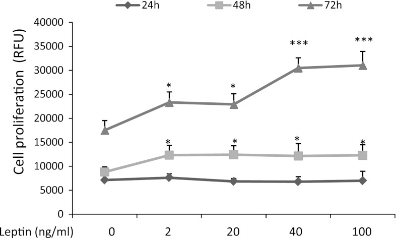
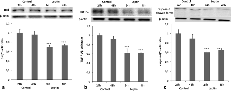
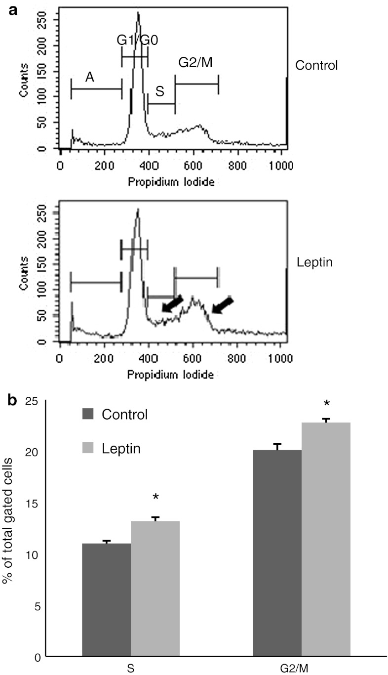
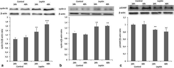
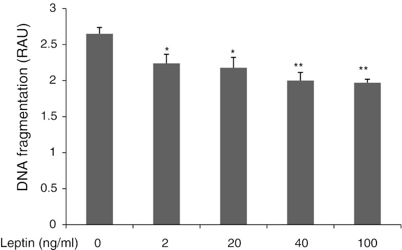
The OVCAR-3 cell line expressing the long (ObRb) and short (ObRt) isoforms of leptin receptor mRNA was used to analyze the effect of leptin on the expression of selected genes and proteins involved in the cell cycle and apoptosis. OVCAR-3 cells were exposed to 2, 20, 40, and 100 ng/ml of leptin. Cell proliferation was determined using the alamarBlue cell viability test and flow cytometry. Apoptosis was measured using a cellular DNA fragmentation ELISA kit. The expression of selected cell cycle and apoptosis genes was evaluated by real-time PCR and confirmed by western blot. The stimulatory action of leptin on cell proliferation was observed as an increase in cells in the S and G2/M phases. Up-regulation of genes responsible for inducing cell proliferation and suppression of genes responsible for inhibition of proliferation were noted. Western blots revealed increased expression of cyclins D and A and inhibition of p21WAF1/CIP1 protein expression by leptin. Inhibition of DNA fragmentation was observed under all leptin doses. Suppression of genes involved in the extrinsic and intrinsic apoptotic pathway was observed. Western blots illustrated decreased Bad, TNFR1, and caspase 6 protein expression in response to leptin treatment. Leptin promotes ovarian cancer cell line growth by up-regulating genes and proteins responsible for inducing cell proliferation as well as down-regulating pro-apoptotic genes and proteins in apoptotic pathways. Results of this study warrant examining the relationship between the risk of ovarian cancer and elevated leptin levels in obese women.
OVCAR-3 细胞系表达瘦素受体 mRNA 的长(ObRb)和短(ObRt)异构体,用于分析瘦素对细胞周期和细胞凋亡相关基因和蛋白表达的影响。将 OVCAR-3 细胞暴露于 2、20、40 和 100ng/ml 的瘦素中。使用 alamarBlue 细胞活力检测法和流式细胞术测定细胞增殖。通过细胞 DNA 片段化 ELISA 试剂盒测定细胞凋亡。通过实时 PCR 评估选定的细胞周期和凋亡基因的表达,并通过 Western blot 进行确认。瘦素对细胞增殖的刺激作用表现为 S 和 G2/M 期细胞增加。注意到与诱导细胞增殖相关的基因上调和与增殖抑制相关的基因下调。Western blot 显示瘦素增加了周期蛋白 D 和 A 的表达,并抑制了 p21WAF1/CIP1 蛋白的表达。在所有瘦素剂量下均观察到 DNA 片段化的抑制。观察到外源性和内源性凋亡途径中相关基因的抑制。Western blot 表明,瘦素处理后 Bad、TNFR1 和 caspase 6 蛋白表达减少。瘦素通过上调与诱导细胞增殖相关的基因和蛋白,下调凋亡途径中的促凋亡基因和蛋白,促进卵巢癌细胞系的生长。本研究的结果表明,需要研究肥胖女性中卵巢癌风险与瘦素水平升高之间的关系。