Wang Honglan, Bonilla Ingrid M, Huang Xin, He Quanhua, Kohr Mark J, Carnes Cynthia A, Ziolo Mark T
Department of Physiology and Cell Biology, The Dorothy M. Davis Heart and Lung Research Institute, The Ohio State University, Columbus, OH 43221, USA.
J Signal Transduct. 2012;2012:645721. doi: 10.1155/2012/645721. Epub 2012 Aug 28.
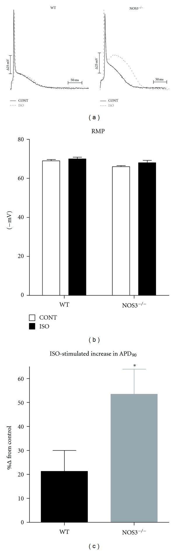
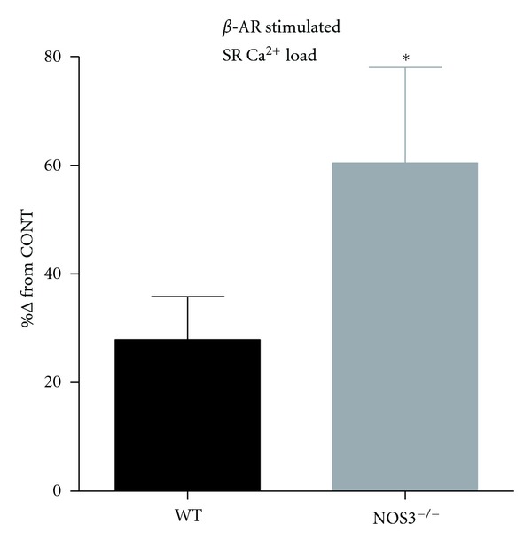
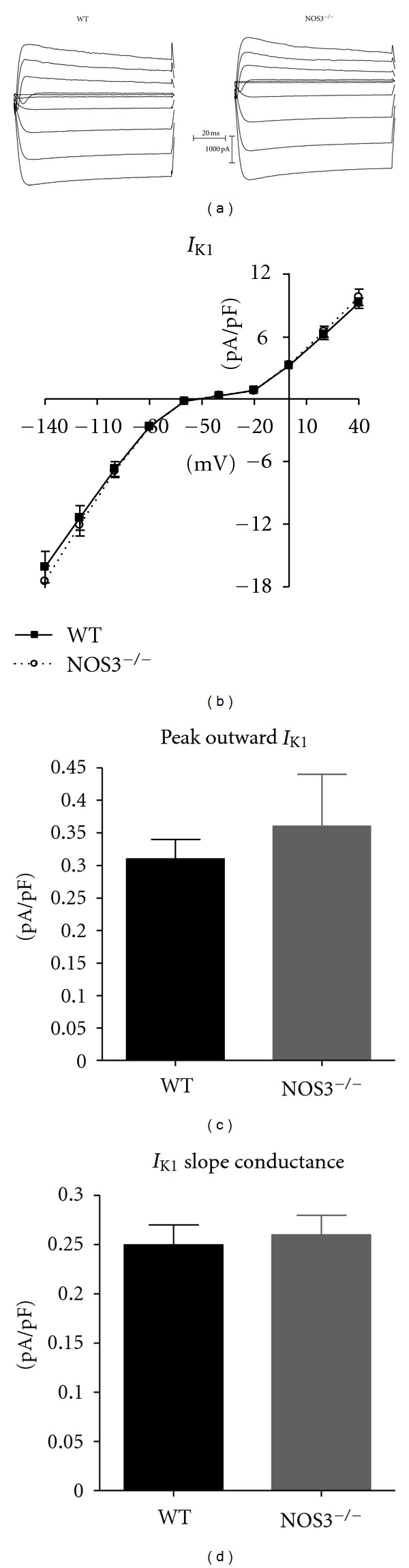
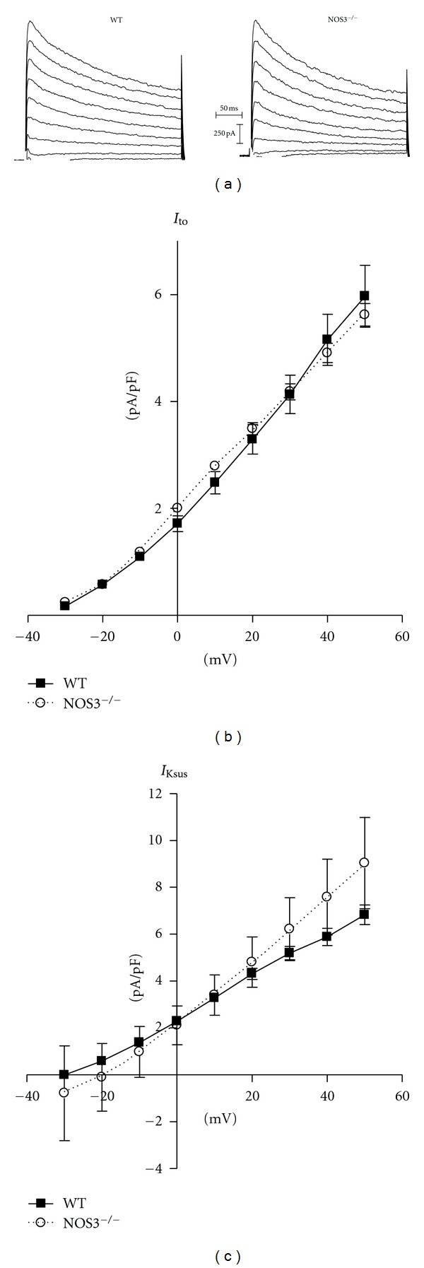
Ventricular myocytes deficient in endothelial nitric oxide synthase (NOS3(-/-)) exhibit prolonged action potential (AP) duration and enhanced spontaneous activity (early and delayed afterdepolarizations) during β-adrenergic (β-AR) stimulation. Studies have shown that nitric oxide is able to regulate various K(+) channels. Our objective was to examine if NOS3(-/-) myocytes had altered K(+) currents. APs, transient outward (I(to)), sustained (I(Ksus)), and inward rectifier (I(K1)) K(+) currents were measured in NOS3(-/-) and wild-type (WT) myocytes. During β-AR stimulation, AP duration (measured as 90% repolarization-APD(90)) was prolonged in NOS3(-/-) compared to WT myocytes. Nevertheless, we did not observe differences in I(to), I(Ksus), or I(K1) between WT and NOS3(-/-) myocytes. Our previous work showed that NOS3(-/-) myocytes had a greater Ca(2+) influx via L-type Ca(2+) channels with β-AR stimulation. Thus, we measured β-AR-stimulated SR Ca(2+) load and found a greater increase in NOS3(-/-) versus WT myocytes. Hence, our data suggest that the prolonged AP in NOS3(-/-) myocytes is not due to changes in I(to), I(Ksus), or I(K1). Furthermore, the increase in spontaneous activity in NOS3(-/-) myocytes may be due to a greater increase in SR Ca(2+) load. This may have important implications for heart failure patients, where arrhythmias are increased and NOS3 expression is decreased.
内皮型一氧化氮合酶缺乏(NOS3(-/-))的心室肌细胞在β-肾上腺素能(β-AR)刺激期间表现出动作电位(AP)时程延长和自发活动增强(早期和延迟后去极化)。研究表明,一氧化氮能够调节各种钾离子通道。我们的目的是研究NOS3(-/-)肌细胞的钾离子电流是否发生改变。在NOS3(-/-)和野生型(WT)肌细胞中测量了动作电位、瞬时外向(I(to))、持续(I(Ksus))和内向整流(I(K1))钾离子电流。在β-AR刺激期间,与WT肌细胞相比,NOS3(-/-)肌细胞的动作电位时程(以90%复极化-APD(90)测量)延长。然而,我们未观察到WT和NOS3(-/-)肌细胞在I(to)、I(Ksus)或I(K1)方面存在差异。我们之前的工作表明,在β-AR刺激下,NOS3(-/-)肌细胞通过L型钙通道有更大的钙内流。因此,我们测量了β-AR刺激下的肌浆网钙负荷,发现与WT肌细胞相比,NOS3(-/-)肌细胞的增加幅度更大。因此,我们的数据表明,NOS3(-/-)肌细胞中动作电位延长并非由于I(to)、I(Ksus)或I(K1)的变化。此外,NOS3(-/-)肌细胞中自发活动的增加可能是由于肌浆网钙负荷的更大增加。这可能对心力衰竭患者具有重要意义,这些患者心律失常增加且NOS3表达降低。