• 文献检索
  • 文档翻译
  • 深度研究
  • 学术资讯
  • Suppr Zotero 插件Zotero 插件
  • 邀请有礼
  • 套餐&价格
  • 历史记录
应用&插件
Suppr Zotero 插件Zotero 插件浏览器插件Mac 客户端Windows 客户端微信小程序
定价
高级版会员购买积分包购买API积分包
服务
文献检索文档翻译深度研究API 文档MCP 服务
关于我们
关于 Suppr公司介绍联系我们用户协议隐私条款
关注我们

Suppr 超能文献

核心技术专利:CN118964589B侵权必究
粤ICP备2023148730 号-1Suppr @ 2026

文献检索

告别复杂PubMed语法,用中文像聊天一样搜索,搜遍4000万医学文献。AI智能推荐,让科研检索更轻松。

立即免费搜索

文件翻译

保留排版,准确专业,支持PDF/Word/PPT等文件格式,支持 12+语言互译。

免费翻译文档

深度研究

AI帮你快速写综述,25分钟生成高质量综述,智能提取关键信息,辅助科研写作。

立即免费体验

正电子发射断层扫描示踪剂在乳腺癌临床成像中的应用。

PET Tracers for Clinical Imaging of Breast Cancer.

机构信息

Department of Nuclear Medicine, University Clinic of Navarra, Avenida Pío XII 36, 31008 Pamplona, Spain.

出版信息

J Oncol. 2012;2012:710561. doi: 10.1155/2012/710561. Epub 2012 Aug 29.

DOI:10.1155/2012/710561
PMID:22973310
原文链接:https://pmc.ncbi.nlm.nih.gov/articles/PMC3437678/
Abstract

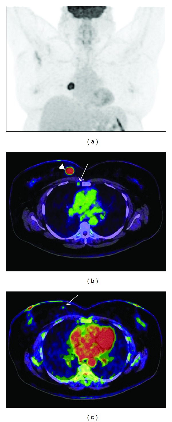
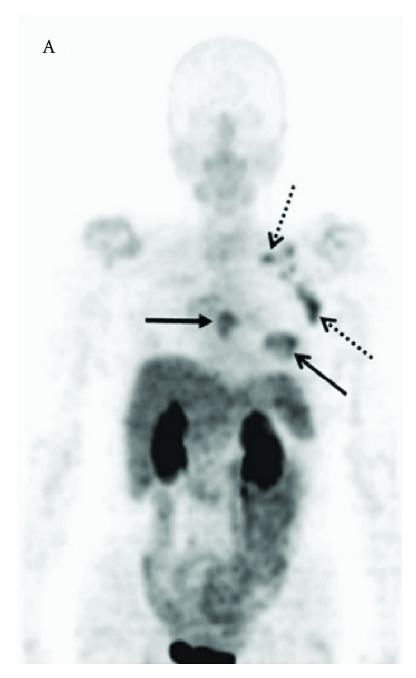
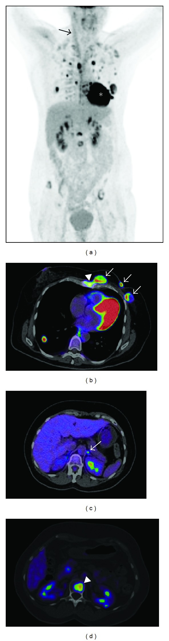
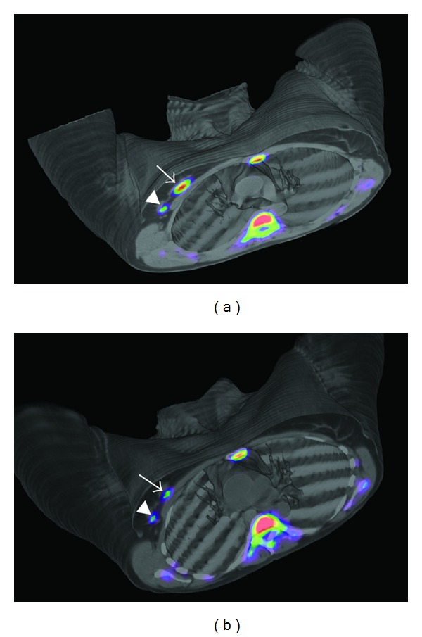
Molecular imaging of breast cancer has undoubtedly permitted a substantial development of the overall diagnostic accuracy of this malignancy in the last years. Accurate tumour staging, design of individually suited therapies, response evaluation, early detection of recurrence and distant lesions have also evolved in parallel with the development of novel molecular imaging approaches. In this context, positron emission tomography (PET) can be probably seen as the most interesting molecular imaging technology with straightforward clinical application for such purposes. Dozens of radiotracers for PET imaging of breast cancer have been tested in laboratory animals. However, in this review we shall focus mainly in the smaller group of PET radiopharmaceuticals that have lead through into the clinical setting. PET imaging can be used to target general metabolic phenomena related to tumoural transformation, including glucose metabolism and cell proliferation, but can also be directed to specific hormone receptors that are characteristic of the breast cancer cell. Many other receptors and transport molecules present in the tumour cells could also be of interest for imaging. Furthermore, molecules related with the tumour microenvironment, tumour induced angiogenesis or even hypoxia could also be used as molecular biomarkers for breast cancer imaging.

摘要

近年来,分子成像技术无疑使乳腺癌的整体诊断准确性得到了显著提高。随着新型分子成像方法的发展,肿瘤分期、个体化治疗方案设计、疗效评估、早期复发和远处转移病灶的检测也取得了进展。在这方面,正电子发射断层扫描(PET)可能是最有趣的分子成像技术,可直接用于这些目的。已经在实验动物中测试了数十种用于乳腺癌 PET 成像的放射性示踪剂。然而,在这篇综述中,我们主要关注的是已经进入临床应用的较小一组 PET 放射性药物。PET 成像可用于靶向与肿瘤转化相关的一般代谢现象,包括葡萄糖代谢和细胞增殖,但也可用于针对乳腺癌细胞特有的特定激素受体。肿瘤细胞中存在的许多其他受体和转运分子也可能对成像感兴趣。此外,与肿瘤微环境、肿瘤诱导的血管生成甚至缺氧相关的分子也可以作为乳腺癌成像的分子生物标志物。

https://cdn.ncbi.nlm.nih.gov/pmc/blobs/49e4/3437678/e03d40f16583/JO2012-710561.005.jpg
https://cdn.ncbi.nlm.nih.gov/pmc/blobs/49e4/3437678/39f18257d933/JO2012-710561.001.jpg
https://cdn.ncbi.nlm.nih.gov/pmc/blobs/49e4/3437678/c3b20c44836f/JO2012-710561.002.jpg
https://cdn.ncbi.nlm.nih.gov/pmc/blobs/49e4/3437678/c46a83c1640a/JO2012-710561.003.jpg
https://cdn.ncbi.nlm.nih.gov/pmc/blobs/49e4/3437678/17d1c40d8767/JO2012-710561.004.jpg
https://cdn.ncbi.nlm.nih.gov/pmc/blobs/49e4/3437678/e03d40f16583/JO2012-710561.005.jpg

相似文献

1
PET Tracers for Clinical Imaging of Breast Cancer.正电子发射断层扫描示踪剂在乳腺癌临床成像中的应用。
J Oncol. 2012;2012:710561. doi: 10.1155/2012/710561. Epub 2012 Aug 29.
2
More advantages in detecting bone and soft tissue metastases from prostate cancer using F-PSMA PET/CT.使用F-PSMA PET/CT检测前列腺癌骨和软组织转移方面有更多优势。
Hell J Nucl Med. 2019 Jan-Apr;22(1):6-9. doi: 10.1967/s002449910952. Epub 2019 Mar 7.
3
New Clinical Indications for (18)F/(11)C-choline, New Tracers for Positron Emission Tomography and a Promising Hybrid Device for Prostate Cancer Staging: A Systematic Review of the Literature.(18)F/(11)C-胆碱的新临床适应证、正电子发射断层扫描的新型示踪剂和用于前列腺癌分期的有前途的混合设备:文献系统评价。
Eur Urol. 2016 Jul;70(1):161-175. doi: 10.1016/j.eururo.2016.01.029. Epub 2016 Feb 2.
4
Position of nuclear medicine modalities in the diagnostic work-up of breast cancer.核医学模式在乳腺癌诊断检查中的地位。
Q J Nucl Med Mol Imaging. 2004 Jun;48(2):109-18.
5
Positron emission tomography in diagnosis and management of invasive breast cancer: current status and future perspectives.正电子发射断层扫描在浸润性乳腺癌诊断与管理中的应用:现状与未来展望
Clin Breast Cancer. 2003 Apr;4 Suppl 1:S55-63. doi: 10.3816/cbc.2003.s.016.
6
Current applications of PET imaging of sex hormone receptors with a fluorinated analogue of estradiol or of testosterone.使用雌二醇或睾酮的氟化类似物对性激素受体进行PET成像的当前应用。
Q J Nucl Med Mol Imaging. 2015 Mar;59(1):4-17. Epub 2015 Feb 19.
7
New PET radiopharmaceuticals beyond FDG for brain tumor imaging.用于脑肿瘤成像的、除氟代脱氧葡萄糖(FDG)之外的新型正电子发射断层显像(PET)放射性药物。
Q J Nucl Med Mol Imaging. 2012 Apr;56(2):173-90.
8
Molecular imaging of pulmonary cancer and inflammation.肺癌与炎症的分子成像
Proc Am Thorac Soc. 2009 Aug 15;6(5):464-8. doi: 10.1513/pats.200902-005AW.
9
Molecular positron emission tomography and PET/CT imaging in urological malignancies.分子正电子发射断层扫描及PET/CT成像在泌尿系统恶性肿瘤中的应用
Eur Urol. 2007 Jun;51(6):1511-20; discussion 1520-1. doi: 10.1016/j.eururo.2007.01.061. Epub 2007 Jan 23.
10
Opportunities for PET to deliver clinical benefit in cancer: breast cancer as a paradigm.正电子发射断层扫描(PET)在癌症临床获益方面的机遇:以乳腺癌为例。
Cancer Imaging. 2010 Jul 6;10(1):144-52. doi: 10.1102/1470-7330.2010.0020.

引用本文的文献

1
[Molecular breast imaging : Positron emission tomography/magnetic resonance imaging and targeted tracers].[分子乳腺成像:正电子发射断层扫描/磁共振成像及靶向示踪剂]
Radiologie (Heidelb). 2025 Mar;65(3):170-177. doi: 10.1007/s00117-024-01403-z. Epub 2025 Feb 3.
2
Characterization of accurate 3D collimator-detector response function for single- and multi-lofthole collimated SPECT cameras.用于单孔和多孔准直 SPECT 相机的精确 3D 准直器-探测器响应函数的特性描述。
Jpn J Radiol. 2024 Nov;42(11):1330-1341. doi: 10.1007/s11604-024-01624-1. Epub 2024 Jul 2.
3
The Role of Molecular Imaging in Personalized Medicine.

本文引用的文献

1
Antibody therapy of cancer.癌症的抗体治疗。
Nat Rev Cancer. 2012 Mar 22;12(4):278-87. doi: 10.1038/nrc3236.
2
Validity of simplified 3'-deoxy-3'-[18F]fluorothymidine uptake measures for monitoring response to chemotherapy in locally advanced breast cancer.简化的 3'-去氧-3'-[18F]氟胸苷摄取测量用于监测局部晚期乳腺癌化疗反应的有效性。
Mol Imaging Biol. 2012 Dec;14(6):777-82. doi: 10.1007/s11307-012-0547-1.
3
Assessment of progesterone receptors in breast carcinoma by PET with 21-18F-fluoro-16α,17α-[(R)-(1'-α-furylmethylidene)dioxy]-19-norpregn-4-ene-3,20-dione.
分子成像在个性化医疗中的作用。
J Pers Med. 2023 Feb 19;13(2):369. doi: 10.3390/jpm13020369.
4
Breast PET/MRI Hybrid Imaging and Targeted Tracers.乳腺 PET/MRI 融合成像与靶向示踪剂。
J Magn Reson Imaging. 2023 Feb;57(2):370-386. doi: 10.1002/jmri.28431. Epub 2022 Sep 27.
5
Non-conventional and Investigational PET Radiotracers for Breast Cancer: A Systematic Review.用于乳腺癌的非传统及研究性正电子发射断层显像(PET)放射性示踪剂:一项系统评价
Front Med (Lausanne). 2022 Apr 12;9:881551. doi: 10.3389/fmed.2022.881551. eCollection 2022.
6
What's in a node? The clinical and radiologic significance of Virchow's node.淋巴结里有什么?维尔肖淋巴结的临床及放射学意义。
Abdom Radiol (NY). 2022 Jun;47(6):2244-2253. doi: 10.1007/s00261-022-03487-4. Epub 2022 Mar 22.
7
Sugar alcohol provides imaging contrast in cancer detection.糖醇为癌症检测提供成像对比。
Sci Rep. 2019 Jul 31;9(1):11092. doi: 10.1038/s41598-019-47275-5.
8
Feasibility study of a point-of-care positron emission tomography system with interactive imaging capability.交互式成像性能的即时检测正电子发射断层成像系统的可行性研究。
Med Phys. 2019 Apr;46(4):1798-1813. doi: 10.1002/mp.13397. Epub 2019 Feb 14.
9
Integrated landscape of copy number variation and RNA expression associated with nodal metastasis in invasive ductal breast carcinoma.浸润性导管癌中与淋巴结转移相关的拷贝数变异和RNA表达的综合图谱。
Oncotarget. 2018 Dec 7;9(96):36836-36848. doi: 10.18632/oncotarget.26386.
10
Molecular imaging of breast cancer: present and future directions.乳腺癌的分子影像学:现状与未来方向。
Front Chem. 2014 Dec 18;2:112. doi: 10.3389/fchem.2014.00112. eCollection 2014.
采用 21-18F-氟-16α,17α-[(R)-(1'-α-糠叉基)二氧]-19-去甲孕甾-4-烯-3,20-二酮进行乳腺癌孕激素受体的 PET 评估。
J Nucl Med. 2012 Mar;53(3):363-70. doi: 10.2967/jnumed.111.098319. Epub 2012 Feb 13.
4
Antiangiogenic agents increase breast cancer stem cells via the generation of tumor hypoxia.抗血管生成药物通过产生肿瘤缺氧增加乳腺癌干细胞。
Proc Natl Acad Sci U S A. 2012 Feb 21;109(8):2784-9. doi: 10.1073/pnas.1018866109. Epub 2012 Jan 23.
5
PET imaging of estrogen receptors as a diagnostic tool for breast cancer patients presenting with a clinical dilemma.正电子发射断层扫描(PET)成像技术作为一种诊断工具,用于诊断在临床上存在两难困境的乳腺癌患者的雌激素受体。
J Nucl Med. 2012 Feb;53(2):182-90. doi: 10.2967/jnumed.111.092734. Epub 2012 Jan 12.
6
Zirconium-89-trastuzumab positron emission tomography as a tool to solve a clinical dilemma in a patient with breast cancer.锆-89-曲妥珠单抗正电子发射断层扫描作为解决一名乳腺癌患者临床困境的工具。
J Clin Oncol. 2012 Feb 20;30(6):e74-5. doi: 10.1200/JCO.2011.38.0204. Epub 2011 Dec 27.
7
Radiosynthesis of [13N]dantrolene, a positron emission tomography probe for breast cancer resistant protein, using no-carrier-added [13N]ammonia.[13N]丹曲林的放射性合成,一种用于乳腺癌耐药蛋白的正电子发射断层扫描探针,使用无载体添加[13N]氨。
Bioorg Med Chem. 2012 Jan 1;20(1):305-10. doi: 10.1016/j.bmc.2011.10.077. Epub 2011 Nov 4.
8
[18F]-3'Deoxy-3'-fluorothymidine positron emission tomography and breast cancer response to docetaxel.[18F]-3'脱氧-3'氟胸苷正电子发射断层扫描与多西他赛治疗乳腺癌的疗效评估。
Clin Cancer Res. 2011 Dec 15;17(24):7664-72. doi: 10.1158/1078-0432.CCR-11-0783. Epub 2011 Oct 25.
9
68Ga-NODAGA-RGDyK for αvβ3 integrin PET imaging. Preclinical investigation and dosimetry.用于αvβ3整合素PET成像的68Ga-NODAGA-RGDyK:临床前研究与剂量测定
Nuklearmedizin. 2011;50(6):225-33. doi: 10.3413/Nukmed-0416-11-06. Epub 2011 Oct 11.
10
Factors influencing the uptake of 18F-fluoroestradiol in patients with estrogen receptor positive breast cancer.影响雌激素受体阳性乳腺癌患者摄取 18F-氟雌二醇的因素。
Nucl Med Biol. 2011 Oct;38(7):969-78. doi: 10.1016/j.nucmedbio.2011.03.002. Epub 2011 May 5.