Department of Radiology, Research Institute of Radiological Science, Yonsei University College of Medicine, Seoul 120-752, Korea.
Korean J Radiol. 2012 Sep-Oct;13(5):586-93. doi: 10.3348/kjr.2012.13.5.586. Epub 2012 Aug 28.
To retrospectively define which histologic characteristics of small-sized hepatocellular carcinomas (HCCs) are related to atypical dynamic enhancement on multi-detector computed tomography (MDCT) imaging.
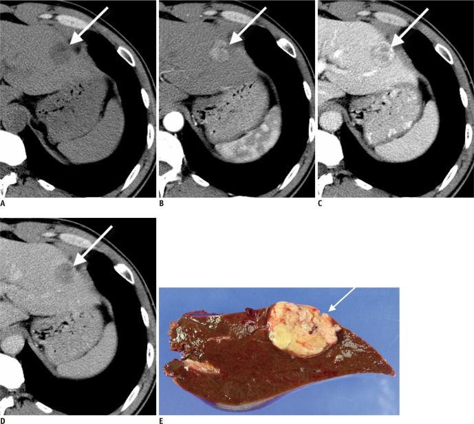
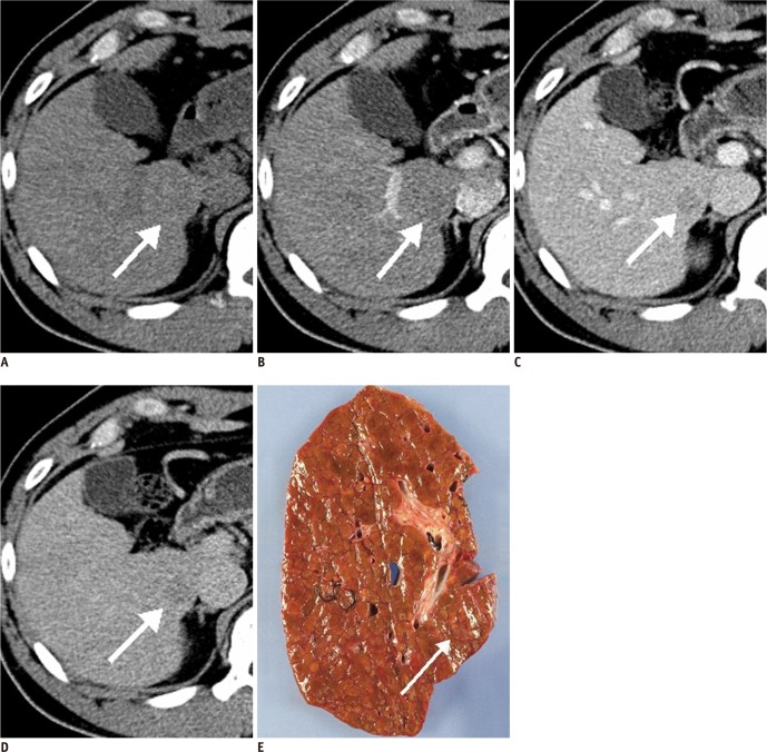
Seventy-three patients with 83 HCCs (3 cm or less in diameter) were included in this study. All patients underwent 4-phase MDCT imaging and subsequent surgery within eight weeks. Two independent radiologists blinded to the histologic findings retrospectively classified the HCCs as either typical (showing increased enhancement on arterial phase images followed by washout in late phase images) or atypical lesions demonstrating any other enhancement pattern. From the original pathologic reports, various histologic characteristics including gross morphology, nuclear histologic grades, presence of capsule formation, and capsule infiltration when a capsule was present, were compared among the two groups.
An atypical enhancement pattern was seen in 30 (36.2%) of the 83 HCCs. The mean size of atypical HCCs (1.71 ± 0.764) was significantly smaller than that of typical HCCs (2.31 ± 0.598, p < 0.001). Atypical HCCs were frequently found to be vaguely nodular in gross morphology (n = 13, 43.3%) and to have grade I nuclear grades (n = 17, 56.7%). Capsule formation was significantly more common in typical HCCs (p < 0.001). Capsular infiltration was also more common in typical HCCs (p = 0.001).
HCCs showing atypical dynamic enhancement on MDCT imaging are usually smaller than typical HCCs, vaguely nodular type in gross morphology in most cases, and well-differentiated in nuclear grades, and they lack of capsule formation or capsular infiltration.
回顾性定义哪些小肝细胞癌(HCC)的组织学特征与多排 CT(MDCT)成像上的非典型动态增强有关。
本研究纳入了 73 例 83 个 HCC(直径 3cm 或以下)患者。所有患者均在 8 周内行 4 期 MDCT 成像及后续手术。两位对组织学发现不知情的独立放射科医生回顾性地将 HCC 分为典型(动脉期增强,晚期廓清)或非典型病变(表现出任何其他增强模式)。从原始病理报告中,比较了两组中各种组织学特征,包括大体形态、核组织学分级、包膜形成的存在以及包膜存在时的包膜浸润。
83 个 HCC 中有 30 个(36.2%)表现为非典型增强模式。非典型 HCC 的平均大小(1.71±0.764)明显小于典型 HCC(2.31±0.598,p<0.001)。非典型 HCC 常表现为大体形态上模糊结节状(n=13,43.3%)和核分级 I 级(n=17,56.7%)。典型 HCC 中包膜形成明显更常见(p<0.001)。包膜浸润在典型 HCC 中也更为常见(p=0.001)。
MDCT 成像上表现为非典型动态增强的 HCC 通常小于典型 HCC,大体形态上多为模糊结节状,核分级多为高分化,缺乏包膜形成或包膜浸润。