Department of Pharmaceutical Chemistry , University of Kansas, Lawrence, KS 66047, USA.
Free Radic Biol Med. 2012 Nov 15;53(10):1877-85. doi: 10.1016/j.freeradbiomed.2012.08.582. Epub 2012 Aug 31.
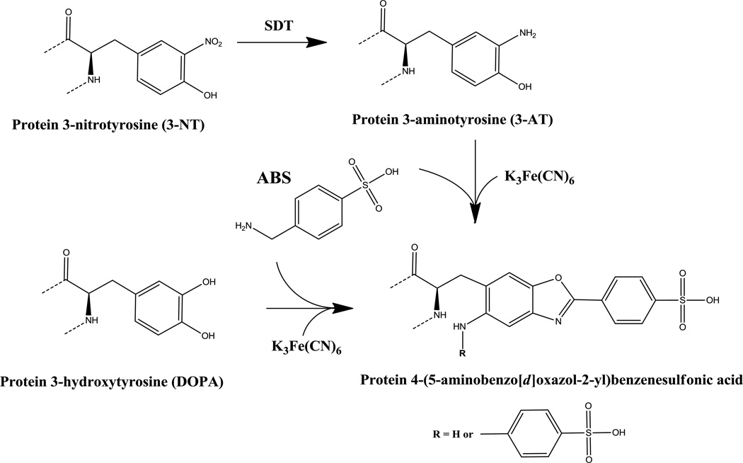
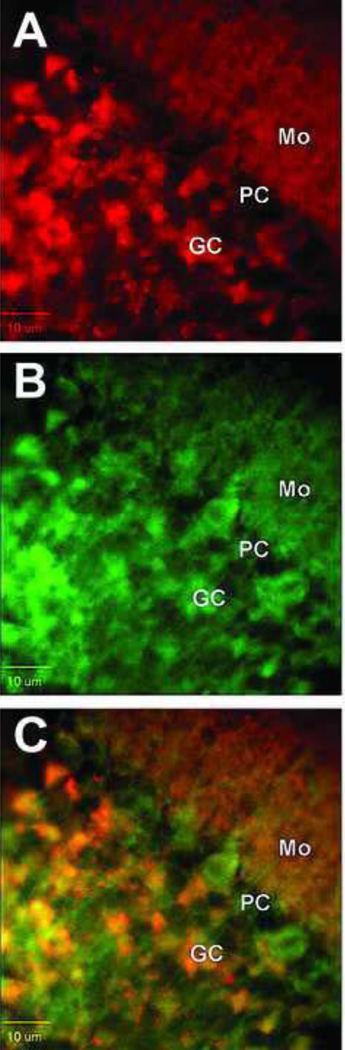
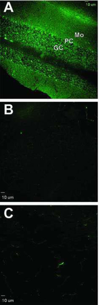
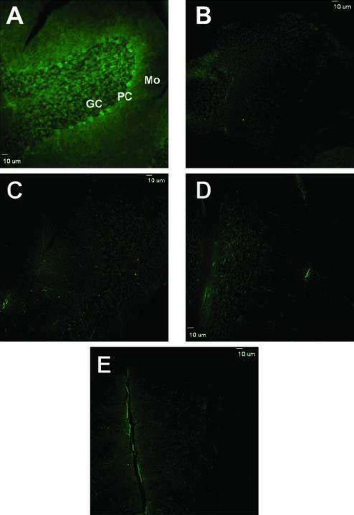
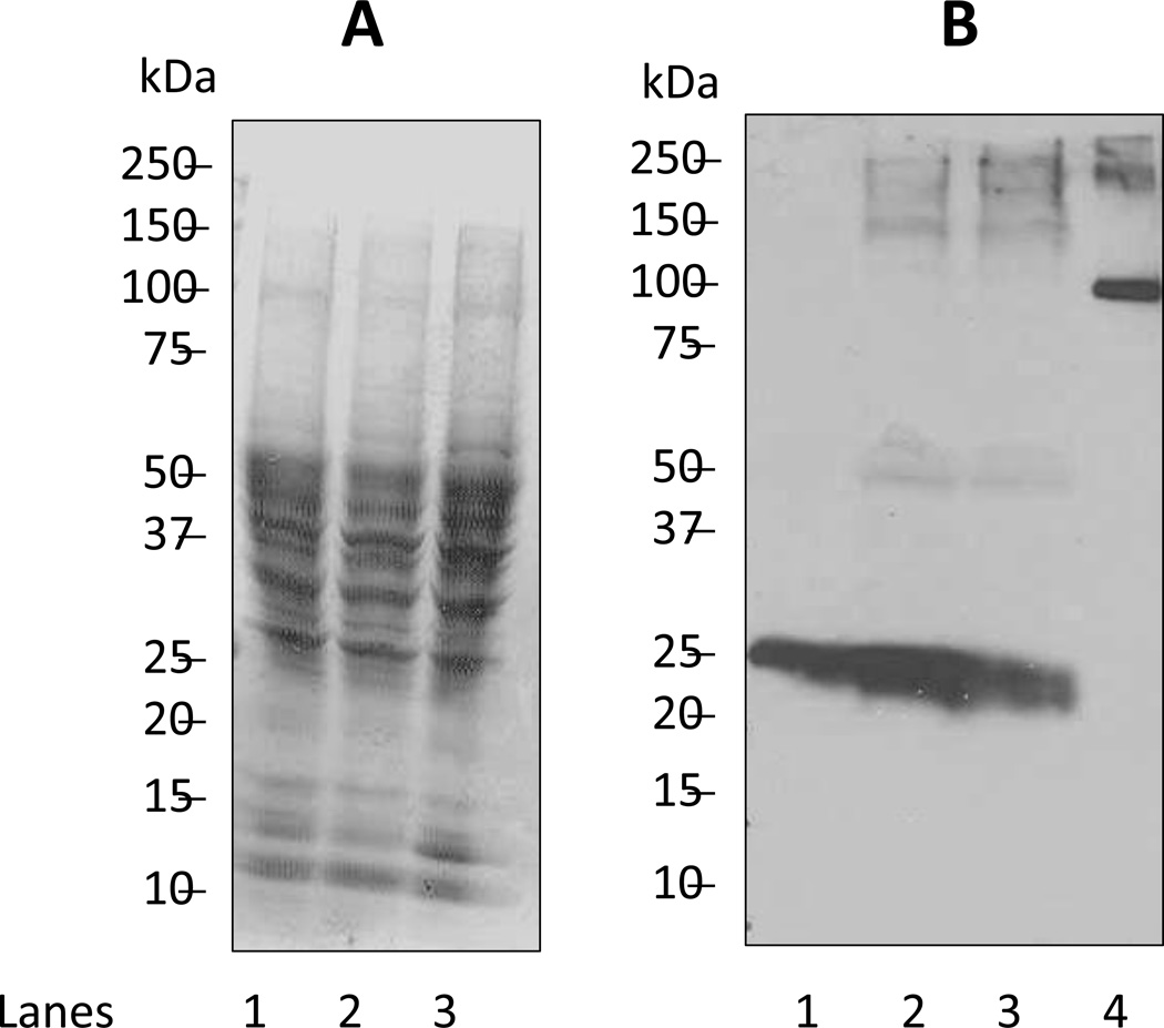
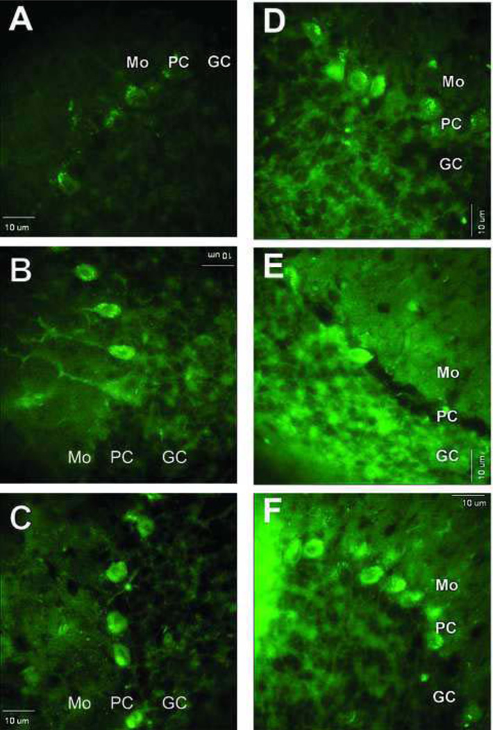
Protein tyrosine nitration is a common biomarker of biological aging and diverse pathologies associated with the excessive formation of reactive oxygen and nitrogen species. Recently, we suggested a novel fluorogenic derivatization procedure for the detection of 3-nitrotyrosine (3-NT) using benzylamine derivatives to convert specifically protein- or peptide-bound 3-NT to a highly fluorescent benzoxazole product. In this study, we applied this procedure to fluorogenic derivatization of protein 3-NT in sections from adult rat cerebellum to: (i) test this method for imaging nitrated proteins in fixed brain tissue sections and (ii) compare the chemical approach to immunohistochemical labeling with anti-3-NT antibodies. Immunofluorescence analysis of cerebellar sections using anti-3-NT antibodies showed differential levels of immunostaining in the molecular, Purkinje, and granule cell layers of the cerebellar cortex; in agreement with previous reports, the Purkinje cells were most highly labeled. Importantly, fluorogenic derivatization reactions of cerebellar proteins with 4-(aminomethyl)benzene sulfonic acid (ABS) and K(3)Fe(CN)(6) at pH 9, after sodium dithionite reduction of 3-NT to 3-aminotyrosine, showed a very similar pattern of relative intensity of cell labeling and improved resolution compared with antibody labeling. Our data demonstrate that ABS derivatization may be either a useful alternative to or a complementary approach to immunolabeling in imaging protein nitration in cells and tissues, including under conditions of dual labeling with antibodies to cell proteins, thus allowing for cellular colocalization of nitrated proteins and any protein of interest.
蛋白质酪氨酸硝化是生物衰老和与活性氧和氮物种过度形成相关的多种病理的常见生物标志物。最近,我们提出了一种使用苄胺衍生物检测 3-硝基酪氨酸(3-NT)的新型荧光衍生化程序,该程序可特异性地将蛋白质或肽结合的 3-NT 转化为高度荧光的苯并恶唑产物。在这项研究中,我们将该程序应用于成年大鼠小脑切片中蛋白质 3-NT 的荧光衍生化,以:(i)测试该方法用于固定脑组织切片中硝化蛋白的成像,(ii)比较化学方法与抗 3-NT 抗体的免疫组织化学标记。使用抗 3-NT 抗体对小脑切片进行免疫荧光分析显示,在小脑皮质的分子层、浦肯野细胞层和颗粒细胞层中,免疫染色水平存在差异;与先前的报道一致,浦肯野细胞的标记水平最高。重要的是,在用 ABS 和 K3Fe(CN)6 进行荧光衍生化反应后,用 4-(氨甲基)苯磺酸(ABS)和 K3Fe(CN)6(pH 9)对小脑蛋白进行衍生化反应,在还原 3-NT 为 3-氨基酪氨酸后,与抗体标记相比,细胞标记的相对强度显示出非常相似的模式,并且分辨率得到了提高。我们的数据表明,在对细胞和组织中的蛋白质硝化进行成像时,ABS 衍生化可能是免疫标记的一种有用替代方法或补充方法,包括在与细胞蛋白的抗体双重标记的情况下,从而允许硝化蛋白和任何感兴趣的蛋白质的细胞共定位。