Mahnken Andreas H
Department of Diagnostic and Interventional Radiology, University Hospital, RWTH Aachen University, Pauwelsstrasse 30, 52074 Aachen, Germany.
ISRN Cardiol. 2012;2012:139823. doi: 10.5402/2012/139823. Epub 2012 Sep 11.
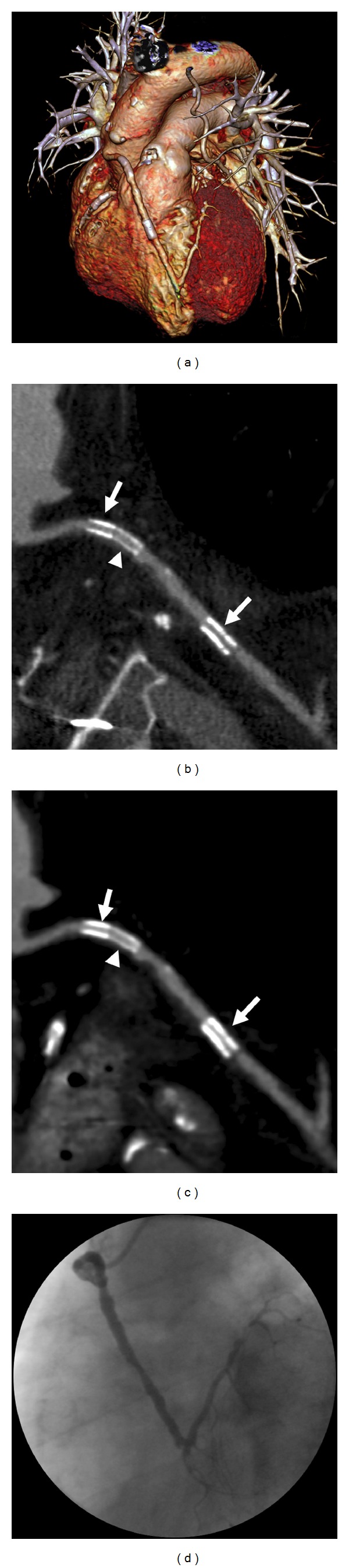
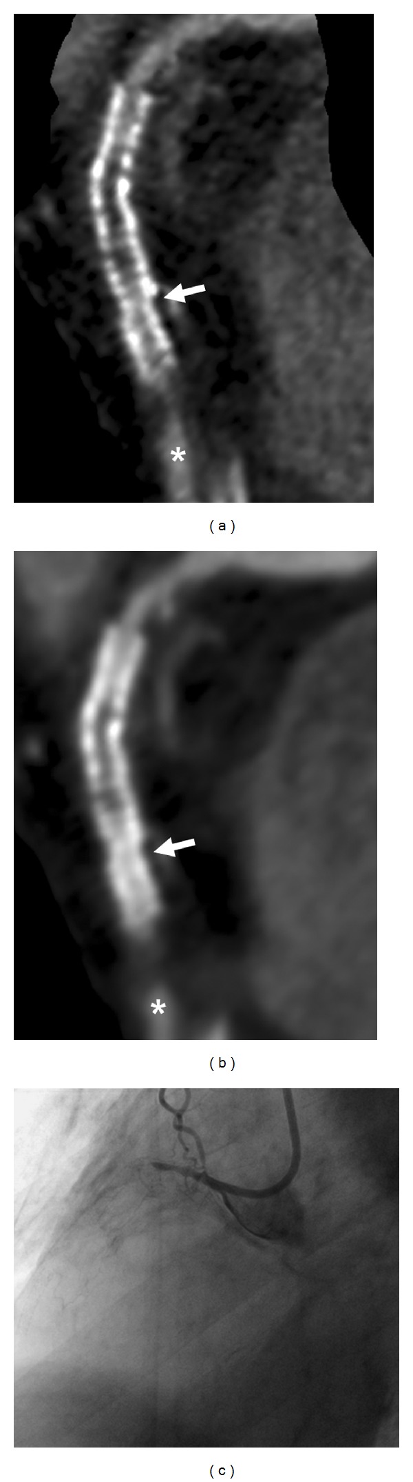
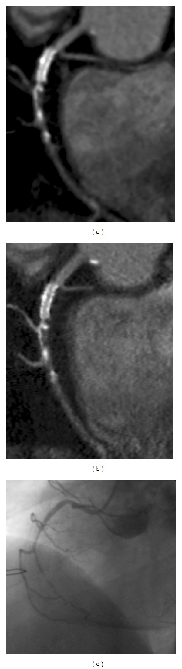
Coronary stenting became a mainstay in coronary revascularization therapy. Despite tremendous advances in therapy, in-stent restenosis (ISR) remains a key problem after coronary stenting. Coronary CT angiography evolved as a valuable tool in the diagnostic workup of patients after coronary revascularization therapy. It has a negative predictive value in the range of 98% for ruling out significant ISR. As CT imaging of coronary stents depends on patient and stent characteristics, patient selection is crucial for success. Ideal candidates have stents with a diameter of 3 mm and more. Nevertheless, even with most recent CT scanners, about 8% of stents are not accessible mostly due to blooming or motion artifacts. While the diagnosis of ISR is currently based on the visual assessment of the stent lumen, functional information on the hemodynamic significance of in-stent stenosis became available with the most recent generation of dual source CT scanners. This paper provides a comprehensive overview on previous developments, current techniques, and clinical evidence for cardiac CT in patients with coronary artery stents.
冠状动脉支架置入术已成为冠状动脉血运重建治疗的主要手段。尽管治疗取得了巨大进展,但支架内再狭窄(ISR)仍是冠状动脉支架置入术后的一个关键问题。冠状动脉CT血管造影已发展成为冠状动脉血运重建治疗后患者诊断检查的一种有价值的工具。其排除显著ISR的阴性预测值在98%左右。由于冠状动脉支架的CT成像取决于患者和支架的特征,因此患者选择对于成功至关重要。理想的候选者应置入直径3毫米及以上的支架。然而,即使使用最新的CT扫描仪,仍有大约8%的支架无法进行评估,这主要是由于伪影或运动伪影所致。虽然目前ISR的诊断基于对支架管腔的视觉评估,但最新一代双源CT扫描仪可提供关于支架内狭窄血流动力学意义的功能信息。本文全面概述了冠状动脉支架置入患者心脏CT的既往进展、当前技术及临床证据。