Department of Medical Pharmacy, Faculty of Pharmacy, Yasuda Women's University, Hiroshima, Japan.
PLoS One. 2012;7(9):e45922. doi: 10.1371/journal.pone.0045922. Epub 2012 Sep 20.
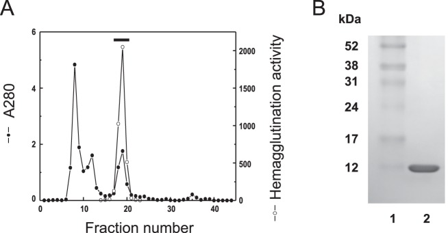
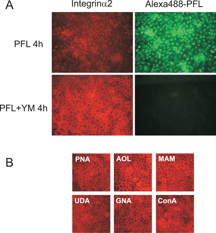
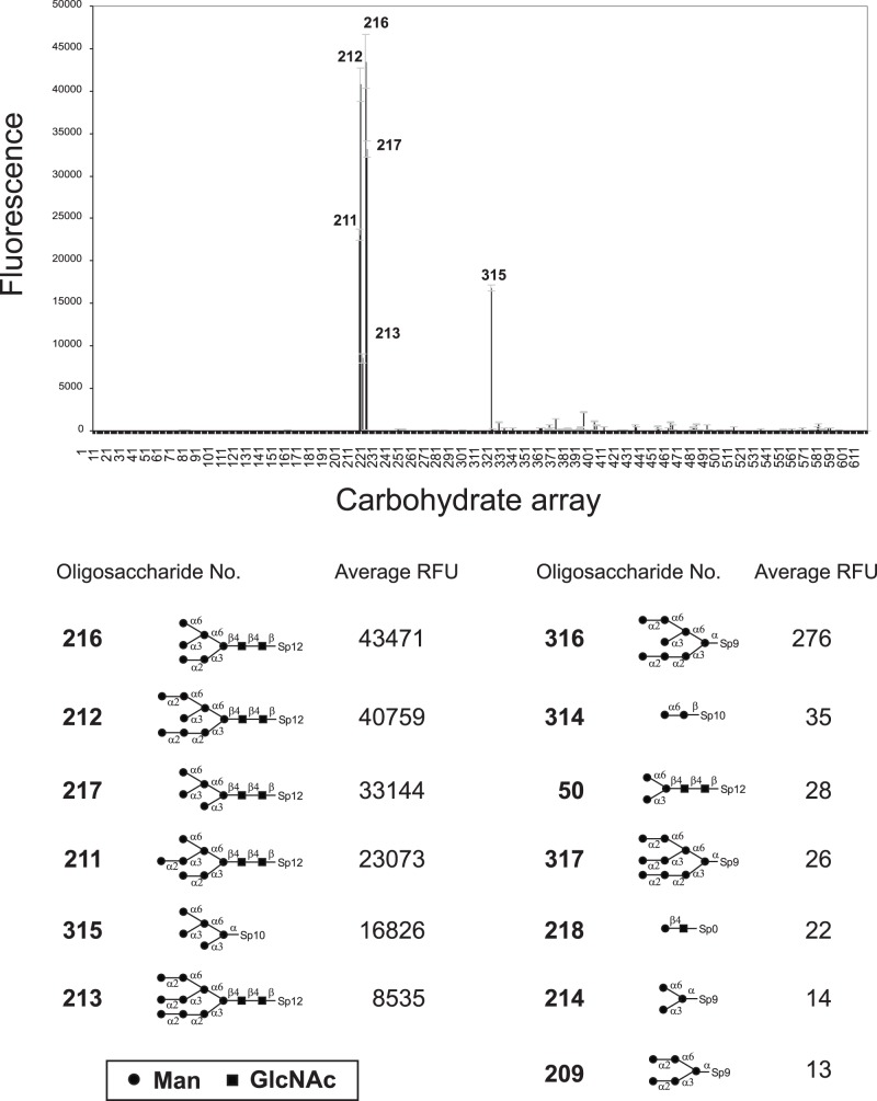
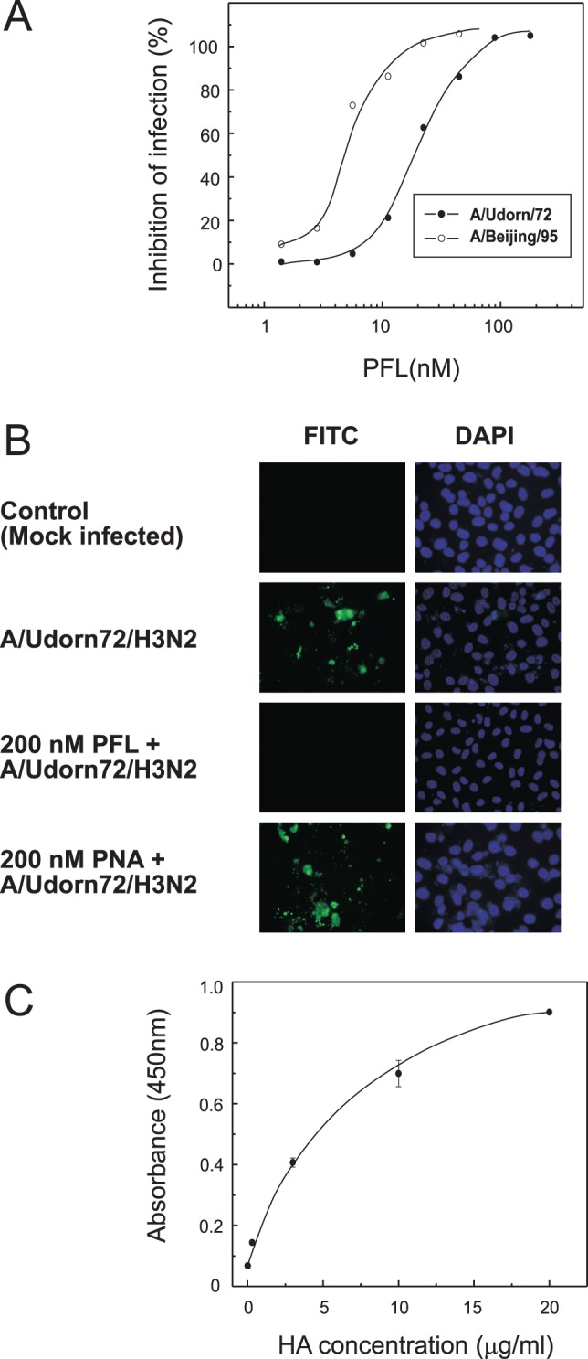
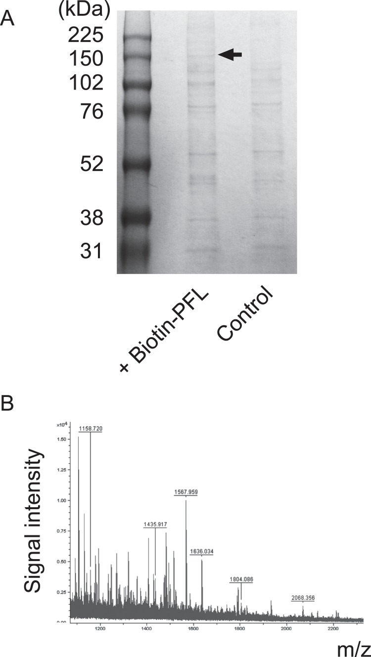
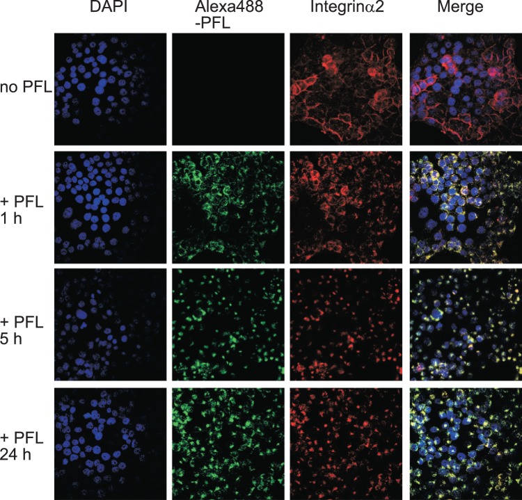
Novel anti-HIV lectin family which shows a strict binding specificity for high mannose glycans has been found in lower organisms. The bacterial orthologue has been identified in the genome of Pseudomonas fluorescens Pf0-1 and the gene coding a putative lectin was cloned, expressed in Escherichia coli and purified by one step gel filtration. Glycan array screening of the recombinant lectin, termed PFL, has revealed that PFL preferentially recognizes high mannose glycans with α1-3 Man that was highly exposed at the D2 position. In contrast, masking of this α1-3 Man with α1-2 Man dramatically impaired lectin-carbohydrate interactions. Reducing terminal disaccharide, GlcNAc-GlcNAc of high mannose glycans was also essential for PFL-binding. PFL showed a potent anti-influenza virus activity by inhibiting the virus entry into cells at doses of low nanomolar concentration. At micromolar concentration or higher, PFL showed a cytotoxicity accompanying loss of the cell adhesion against human gastric cancer MKN28 cells. The cell surface molecule to which PFL bound was co-precipitated with biotin-labeled PFL and identified as integrin α2 by peptide mass fingerprinting using MALDI-TOF mass spectrometry. Intriguingly, upon treatment with exogenous PFL, integrin α2 on the cell surface underwent rapid internalization to the cytoplasm and accumulated to perinuclear region, together with the bound PFL. The resulting loss of cell adherence would trigger a signaling pathway that induced anoikis-like cell death. These events were effectively inhibited by pretreatment of PFL with mannnan, indicating the involvement of high mannose glycans on PFL-induced cell death that was triggered by PFL-integrin α2 interactions.
在较低等生物中发现了一种新型抗 HIV 凝集素家族,该家族对高甘露糖聚糖表现出严格的结合特异性。假单胞菌荧光 Pf0-1 的基因组中已鉴定出细菌同源物,克隆了编码假定凝集素的基因,并在大肠杆菌中表达,然后通过一步凝胶过滤进行纯化。重组凝集素(称为 PFL)的糖基阵列筛选表明,PFL 优先识别高度暴露在 D2 位置的α1-3 Man 的高甘露糖聚糖。相比之下,用α1-2 Man 掩盖这种α1-3 Man 会严重损害凝集素-碳水化合物相互作用。高甘露糖聚糖的还原末端二糖 GlcNAc-GlcNAc 也是 PFL 结合所必需的。PFL 在纳摩尔浓度的低剂量下通过抑制病毒进入细胞来显示出强大的抗流感病毒活性。在微摩尔浓度或更高浓度下,PFL 显示出细胞毒性,同时伴随着人胃癌 MKN28 细胞的细胞黏附丧失。与 PFL 结合的细胞表面分子与生物素标记的 PFL 一起共沉淀,并通过基质辅助激光解吸电离飞行时间质谱法的肽质量指纹图谱鉴定为整合素α2。有趣的是,在用外源性 PFL 处理后,细胞表面上的整合素α2迅速内化到细胞质中,并与结合的 PFL 一起积累到核周区域。由此导致的细胞黏附丧失将触发诱导类似凋亡的细胞死亡的信号通路。这些事件通过用甘露聚糖预先处理 PFL 而有效地被抑制,表明高甘露糖聚糖参与了由 PFL-整合素α2 相互作用引发的 PFL 诱导的细胞死亡。