Department of Pediatrics, Division of Neonatology, Johns Hopkins University-School of Medicine, Baltimore, MD 21287, USA.
J Perinatol. 2013 May;33(5):374-82. doi: 10.1038/jp.2012.124. Epub 2012 Oct 4.
Opioids and clonidine, used in for sedation, analgesia and control of opioid withdrawal in neonates, directly or indirectly activate opioid receptors (OPRs) expressed in immune cells. Therefore, our objective is to study how clinically relevant concentrations of different opioids and clonidine change cytokine levels in cultured whole blood from preterm and full-term infants.
Using blood from preterm (≤ 30 weeks gestational age (GA), n=7) and full-term ( ≥ 37 weeks GA, n=19) infants, we investigated the changes in cytokine profile (IL-1β, IL-6, IL-8, IL-10, IL-12p70 and TNF-α), cyclic adenosine monophosphate (cAMP) levels and μ-, δ- and κ- opioid receptor (OPR) gene and protein expression, following in-vitro exposure to morphine, methadone, fentanyl or clonidine at increasing concentrations ranging from 0 to 1 mM.
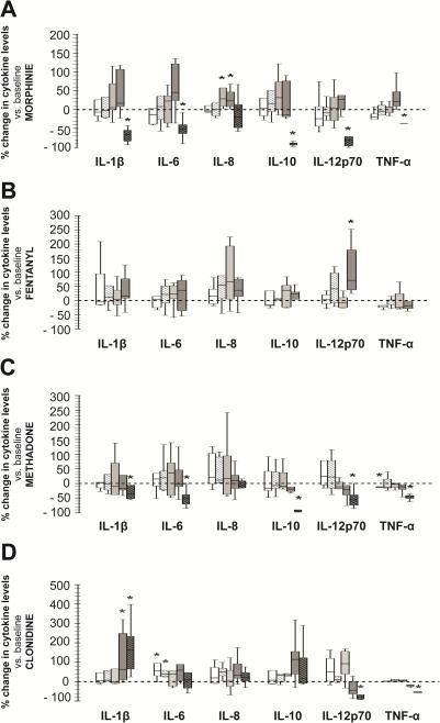
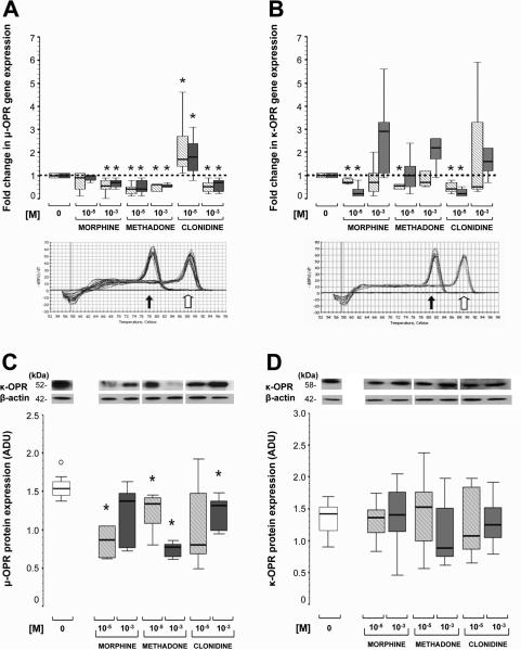
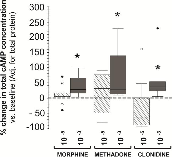
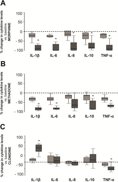
Following lipopolysaccharide activation, IL-10 levels were 146-fold greater in cultured blood from full-term than from preterm infants. Morphine and methadone, but not fentanyl, at >10(-5) M decreased all tested cytokines except IL-8. In contrast, clonidine at <10(-9) M increased IL-6, while at >10(-5) M increased IL-1β and decreased TNF-α levels. All cytokine changes followed the same patterns in preterm and full-term infant cultured blood and matched increases in cAMP levels. All three μ-, δ- and κ-OPR genes were expressed in mononuclear cells (MNC) from preterm and full-term infants. Morphine, methadone and clonidine, but not fentanyl, at >10(-5)M decreased the expression of μ-OPR, but not δ- or κ-OPRs.
Generalized cytokine suppression along with downregulation of μ-OPR expression observed in neonatal MNC exposed to morphine and methadone at clinically relevant concentrations contrast with the modest effects observed with fentanyl and clonidine. Therefore, we speculate that fentanyl and clonidine may be safer therapeutic choices for sedation and control of opioid withdrawal and pain in neonates.
阿片类药物和可乐定用于镇静、镇痛和控制新生儿阿片戒断,它们直接或间接地激活免疫细胞中表达的阿片受体(OPRs)。因此,我们的目标是研究不同阿片类药物和可乐定的临床相关浓度如何改变早产儿和足月儿培养全血中的细胞因子水平。
使用来自早产儿(≤30 周胎龄(GA),n=7)和足月儿(≥37 周 GA,n=19)的血液,我们研究了在体外暴露于不同浓度(0 至 1 mM)的吗啡、美沙酮、芬太尼或可乐定后,细胞因子谱(IL-1β、IL-6、IL-8、IL-10、IL-12p70 和 TNF-α)、环磷酸腺苷(cAMP)水平以及 μ、δ 和 κ-阿片受体(OPR)基因和蛋白表达的变化。
脂多糖激活后,来自足月儿的培养血中 IL-10 水平比早产儿高 146 倍。吗啡和美沙酮(但芬太尼没有)在>10(-5) M 时降低了所有测试的细胞因子,除了 IL-8。相反,可乐定在<10(-9) M 时增加了 IL-6,而在>10(-5) M 时增加了 IL-1β并降低了 TNF-α水平。所有细胞因子变化在早产儿和足月儿培养血中呈现相同模式,并伴有 cAMP 水平的相应增加。所有三种 μ、δ 和 κ-OPR 基因均在早产儿和足月儿的单核细胞(MNC)中表达。吗啡、美沙酮和可乐定(但芬太尼没有)在>10(-5) M 时降低了 μ-OPR 的表达,但没有降低 δ 或 κ-OPR。
与芬太尼和可乐定相比,在临床相关浓度下暴露于吗啡和美沙酮的新生儿 MNC 中观察到的普遍细胞因子抑制以及 μ-OPR 表达下调,这表明芬太尼和可乐定可能是新生儿镇静和控制阿片戒断和疼痛的更安全的治疗选择。