Key Laboratory of Systems Biology Medicine of Jiangxi Province, College of Basic Medical Science, Jiujiang University, Jiujiang, China.
Int J Nanomedicine. 2012;7:5079-90. doi: 10.2147/IJN.S36150. Epub 2012 Sep 21.
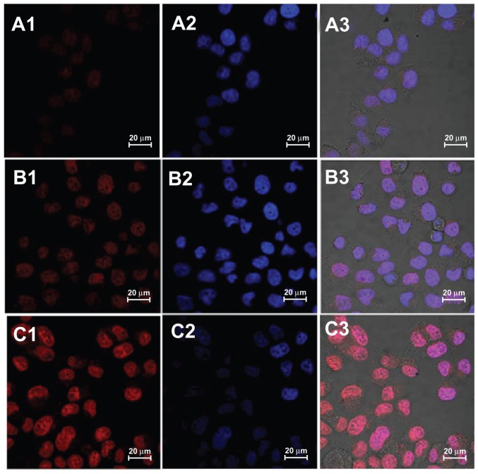
Supramolecular micelles as drug-delivery vehicles are generally unable to enter the nucleus of nondividing cells. In the work reported here, nuclear localization signal (NLS)-modified polymeric micelles were studied with the aim of improving nuclear drug delivery.
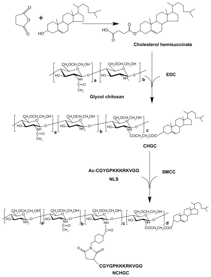
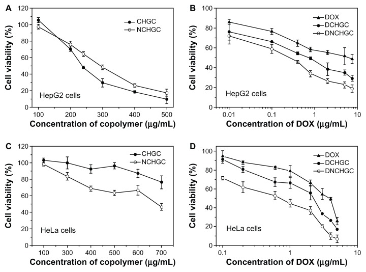
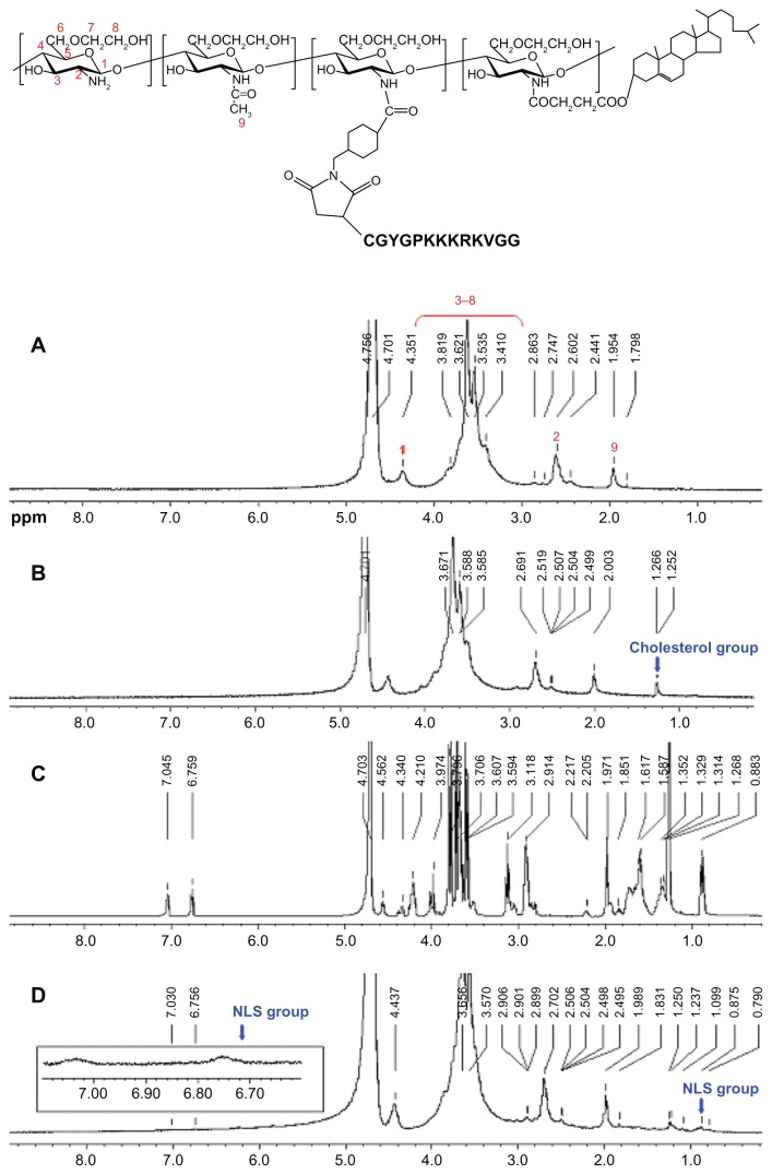
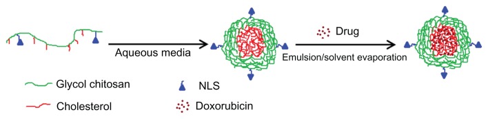
In this research, cholesterol-modified glycol chitosan (CHGC) was synthesized. NLS-conjugated CHGC (NCHGC) was synthesized and characterized using proton nuclear magnetic resonance spectroscopy, dynamic light scattering, and fluorescence spectroscopy. Doxorubicin (DOX), an anticancer drug with an intracellular site of action in the nucleus, was chosen as a model drug. DOX-loaded micelles were prepared by an emulsion/solvent evaporation method. The cellular uptake of different DOX formulations was analyzed by flow cytometry and confocal laser scanning microscopy. The cytotoxicity of blank micelles, free DOX, and DOX-loaded micelles in vitro was investigated by 3-(4,5-dimethylthiazol-2-yl)-2,5-diphenyltetrazolium bromide (MTT) assay in HeLa and HepG2 cells.
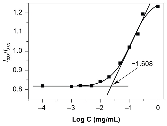
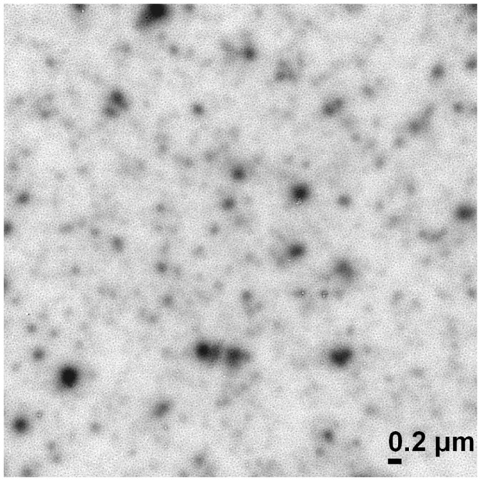
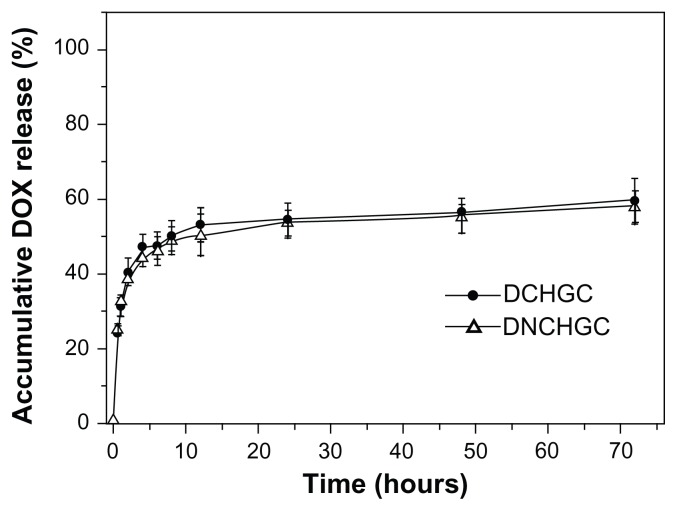
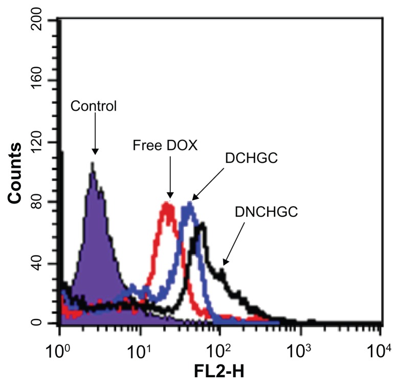
The degree of substitution was 5.9 cholesterol and 3.8 NLS groups per 100 sugar residues of the NCHGC conjugate. The critical aggregation concentration of the NCHGC micelles in aqueous solution was 0.0209 mg/mL. The DOX-loaded NCHGC (DNCHGC) micelles were observed as being almost spherical in shape under transmission electron microscopy, and the size was determined as 248 nm by dynamic light scattering. The DOX-loading content of the DNCHGC micelles was 10.1%. The DOX-loaded micelles showed slow drug-release behavior within 72 hours in vitro. The DNCHGC micelles exhibited greater cellular uptake and higher amounts of DOX in the nuclei of HeLa cells than free DOX and DOX-loaded CHGC (DCHGC) micelles. The half maximal inhibitory concentration (IC(50)) values of free DOX, DCHGC, and DNCHGC micelles against HepG2 cells were 4.063, 0.591, and 0.171 μg/mL, respectively. Moreover, the IC(50) values of free DOX (3.210 μg/mL) and the DCHGC micelles (1.413 μg/mL) against HeLa cells were nearly 6.96- and 3.07-fold (P < 0.01), respectively, higher than the IC(50) value of the DNCHGC micelles (0.461 μg/mL).
The results of this study suggest that novel NCHGC micelles could be a potential carrier for nucleus-targeting delivery.
作为药物递送载体的超分子胶束一般无法进入非分裂细胞的核内。在本研究中,研究了核定位信号(NLS)修饰的聚合物胶束,旨在改善核内药物递送。
本研究合成了胆固醇修饰的乙二醇壳聚糖(CHGC)。采用质子核磁共振波谱、动态光散射和荧光光谱对 NLS 修饰的 CHGC(NCHGC)进行了合成和表征。选择阿霉素(DOX)作为具有核内细胞内作用部位的抗癌药物作为模型药物。通过乳液/溶剂蒸发法制备载 DOX 的胶束。通过流式细胞术和共聚焦激光扫描显微镜分析不同 DOX 制剂的细胞摄取情况。通过 3-(4,5-二甲基噻唑-2-基)-2,5-二苯基四氮唑溴盐(MTT)测定法在 HeLa 和 HepG2 细胞中研究空白胶束、游离 DOX 和载 DOX 胶束的体外细胞毒性。
NCHGC 缀合物的取代度为每 100 个糖残基 5.9 个胆固醇和 3.8 个 NLS 基团。NCHGC 胶束在水溶液中的临界聚集浓度为 0.0209 mg/mL。透射电子显微镜观察到载 DOX 的 NCHGC(DNCHGC)胶束几乎呈球形,动态光散射法测定粒径为 248nm。DNCHGC 胶束的 DOX 载药量为 10.1%。DNCHGC 胶束在体外 72 小时内表现出缓慢的药物释放行为。DNCHGC 胶束在 HeLa 细胞中的细胞摄取和细胞核内 DOX 含量均高于游离 DOX 和载 DOX 的 CHGC(DCHGC)胶束。游离 DOX、DCHGC 和 DNCHGC 胶束对 HepG2 细胞的半数最大抑制浓度(IC(50))值分别为 4.063、0.591 和 0.171μg/mL。此外,游离 DOX(3.210μg/mL)和 DCHGC 胶束(1.413μg/mL)对 HeLa 细胞的 IC(50)值分别比 DNCHGC 胶束(0.461μg/mL)高近 6.96-和 3.07 倍(P<0.01)。
本研究结果表明,新型 NCHGC 胶束可能成为一种有潜力的核靶向递送载体。