Langen H-J, Koehler S, Bielmeier J, Jocher R, Kranzfelder D, Jagusch N, Treutlein G, Wetzler Th, Müller J, Ott G
Department of Radiology, The Medical Mission Clinic, Salvatorstra β e 7, 97074 Wuerzburg, Germany.
Radiol Res Pract. 2012;2012:526293. doi: 10.1155/2012/526293. Epub 2012 Oct 11.
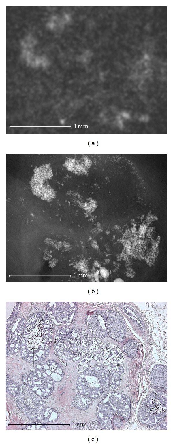
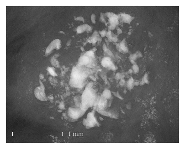
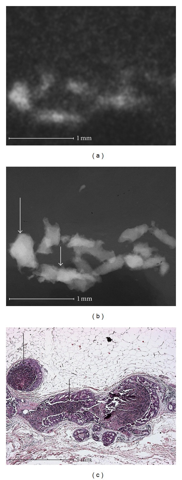
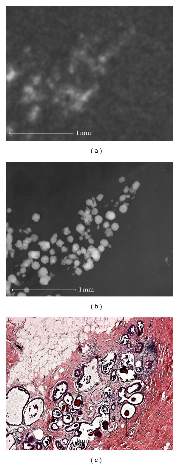
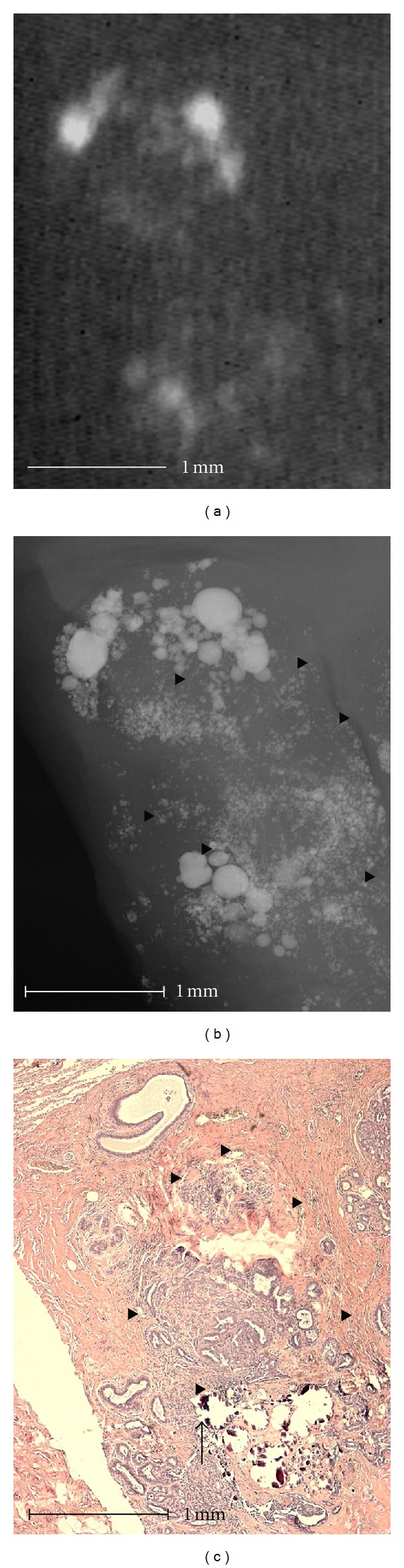
Introduction. Does high-resolution visualization of microcalcifications improve diagnostic reliability? Method. X-rays were taken of mamma specimens with microcalcifications in 32 patients (10 malignant; 22 benign) using conventional radiography (12 Lp/mm) and high-resolution radiography (2000 Lp/mm). Histological sections were subsequently prepared and correlated to the microradiographic image and every calcification was assigned an exact malignant or benign histological diagnosis. Five radiologists classified single groups of calcifications in both methods according to the BIRADS classification system. Results. Using microradiography microcalcifications can be shown in high resolution at the cell level including histological correlation. In some cases, the diagnostic validity was improved by the high resolution in microradiography. In other cases, the high resolution resulted in more visible calcifications, thus giving benign calcifications a malignant appearance. In the BIRADS 2 and 3 group, the probability of malignancy was 28.6% in the conventional radiography evaluation and 37.8% in the microradiography evaluation. In the BIRADS 4 and 5 group, the probability of malignancy was 34.2% in the conventional radiography evaluation and 24.4% in the microradiography evaluation. The differences were not significant. Summary. Overall, the improved resolution in microradiography did not show an improvement in diagnostic accuracy compared to conventional radiography.
引言。微钙化的高分辨率可视化是否能提高诊断可靠性?方法。对32例有微钙化的乳腺标本患者(10例恶性;22例良性)进行X线检查,采用传统放射摄影(12线对/毫米)和高分辨率放射摄影(2000线对/毫米)。随后制备组织学切片,并与微放射图像相关联,对每个钙化灶进行准确的恶性或良性组织学诊断。5名放射科医生根据BIRADS分类系统对两种方法中的单组钙化灶进行分类。结果。使用微放射摄影可以在细胞水平以高分辨率显示微钙化,包括组织学相关性。在某些情况下,微放射摄影的高分辨率提高了诊断有效性。在其他情况下,高分辨率导致钙化更明显,从而使良性钙化呈现恶性外观。在BIRADS 2和3组中,传统放射摄影评估的恶性概率为28.6%,微放射摄影评估为37.8%。在BIRADS 4和5组中,传统放射摄影评估的恶性概率为34.2%,微放射摄影评估为24.4%。差异无统计学意义。总结。总体而言,与传统放射摄影相比,微放射摄影分辨率的提高并未显示出诊断准确性的提高。