Pathology Department, Ain Shams University, Cairo, Egypt.
Diagn Pathol. 2012 Oct 30;7:149. doi: 10.1186/1746-1596-7-149.
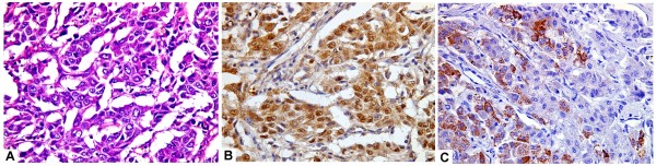
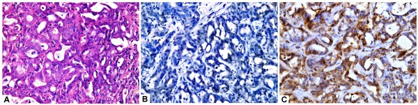
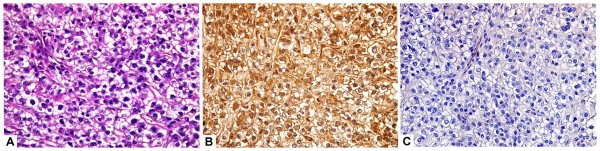
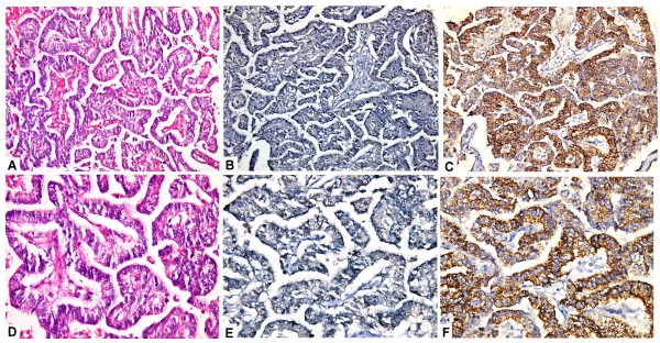
The ability to distinguish hepatocellular carcinoma (HCC) from metastatic carcinoma (MC) involving the liver and cholangiocarcinoma (CC) by immunohistochemistry has been limited by the lack of a reliable positive marker for hepatocellular differentiation. Arginase-1 is a marker for HCC recently described in some literature.
To examine the immunohistochemical staining of arginase-1 in cases of HCC, MC involving the liver and CC as compared to hepatocyte paraffin antigen -1 (HepPar-1) in an attempt to further define the diagnostic utility of arginase-1 in differentiating these tumors.
A comparative immunohistochemical study of arginase-1 and HepPar-1expression was performed in 50 HCC cases, 38 cases of MC to the liver from varying sites, 12 cases of CC and 10 specimens of normal liver tissues. The predictive capacity of arginase-1 and HepPar-1 staining was determined using sensitivity, specificity, positive predictive value, and negative predictive value calculations.
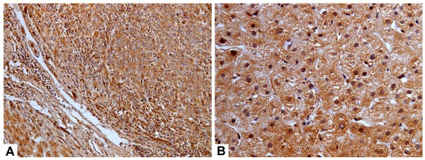
All normal liver tissues (no=10), non- neoplastic cirrhotic liver tissues adjacent to HCC (no=42) as well as those adjacent to MC (no= 9) showed diffuse and strong immunostaining for both arginase-1 and HepPar-1. Arginase-1 demonstrated positive immunoreactivity in 42 of 50 (84%) cases of HCC compared with 35 of 50 (70%) for HepPar-1. Only one of 38 (2.6%) cases of MC and one of 12 (8.3%) cases of CC showed positive immunoreactivity for arginase-1. In contrast, HepPar-1 immunoreactivity was detected in 6 of 38 (15.8%) cases of MC and in 2 of 12 (16.7%) cases of CC. Arginase -1 showed a significantly higher sensitivity for HCC diagnosis (84%) compared to HepPar -1(70%) (p=0.016). The specificity of arginase-1 for HCC diagnosis was higher (96%) than that of HepPar -1 (84%); nevertheless, this was not statistically significant (p=0.109). Howerver, the combination of both immunomarkers for the diagnosis of HCC, raised the specificity to 100%.
Arginase-1 immunostaining has a higher sensitivity and specificity than HepPar-1 for HCC diagnosis. Furthermore, the combined use of arginase-1 and HepPar-1 can provide a potentially promising tool to improve the accuracy in distinguishing HCC from metastatic carcinoma and cholangiocarcinoma.
The virtual slide(s) for this article can be found here: http://www.diagnosticpathology.diagnomx.eu/vs/9991436558072434.
通过免疫组织化学来区分肝癌(HCC)与肝转移癌(MC)和胆管细胞癌(CC)的能力受到缺乏可靠的肝细胞分化阳性标志物的限制。精氨酸酶-1 是最近在一些文献中描述的 HCC 标志物。
通过比较 HCC、来自不同部位的肝 MC 和 CC 中精氨酸酶-1 和 HepPar-1 的免疫组织化学染色,进一步确定精氨酸酶-1 在区分这些肿瘤中的诊断效用。
对 50 例 HCC 病例、38 例来自不同部位的肝 MC、12 例 CC 和 10 例正常肝组织进行了精氨酸酶-1 和 HepPar-1 表达的对比免疫组织化学研究。使用敏感性、特异性、阳性预测值和阴性预测值计算来确定精氨酸酶-1 和 HepPar-1 染色的预测能力。
所有正常肝组织(无=10)、非肿瘤性 HCC 旁肝硬化组织(无=42)以及 MC 旁肝硬化组织(无=9)均显示出精氨酸酶-1 和 HepPar-1 的弥漫性强免疫染色。与 HepPar-1 相比,精氨酸酶-1 在 50 例 HCC 中的 42 例(84%)中显示出阳性免疫反应性,而在 50 例 HCC 中的 35 例(70%)中显示出阳性免疫反应性。只有 1 例(2.6%)MC 和 1 例(8.3%)CC 显示出精氨酸酶-1 的阳性免疫反应性。相比之下,HepPar-1 免疫反应性在 38 例 MC 中的 6 例(15.8%)和 12 例 CC 中的 2 例(16.7%)中被检测到。精氨酸酶-1 对 HCC 诊断的敏感性(84%)明显高于 HepPar-1(70%)(p=0.016)。精氨酸酶-1 对 HCC 诊断的特异性(96%)高于 HepPar-1(84%);然而,这并不具有统计学意义(p=0.109)。然而,两种免疫标志物联合用于 HCC 的诊断,特异性提高到 100%。
精氨酸酶-1 免疫染色对 HCC 的诊断具有比 HepPar-1 更高的敏感性和特异性。此外,精氨酸酶-1 和 HepPar-1 的联合使用可以为提高 HCC 与转移性癌和胆管细胞癌的鉴别准确性提供一种有前途的工具。
本文的虚拟幻灯片可在此处找到:http://www.diagnosticpathology.diagnomx.eu/vs/9991436558072434。