Department of Biology, Center for Research on Biological Clocks, Texas A&M University, College Station, Texas, United States of America.
PLoS One. 2012;7(11):e49555. doi: 10.1371/journal.pone.0049555. Epub 2012 Nov 14.
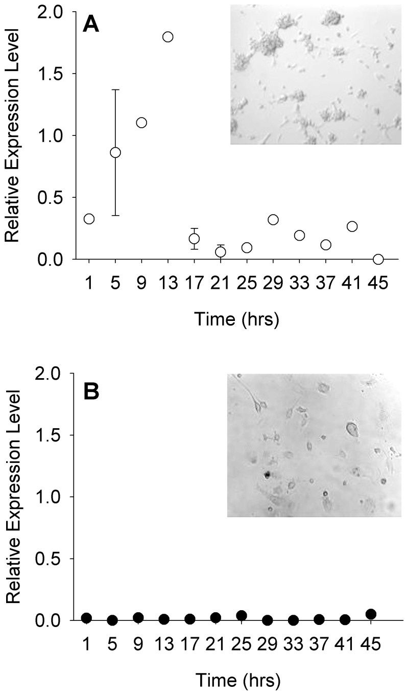
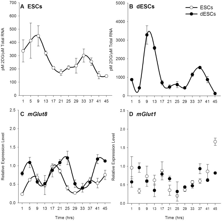
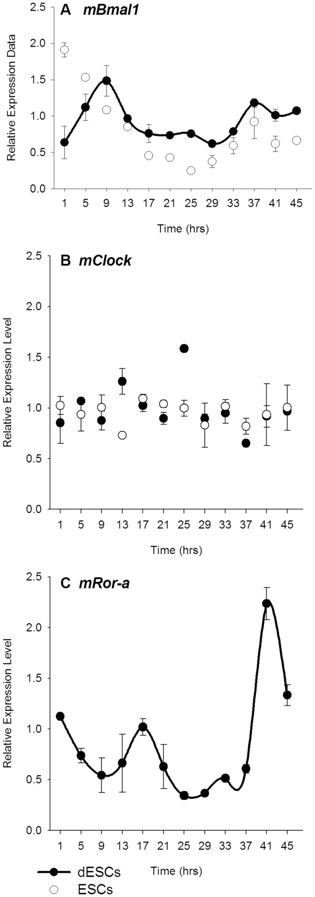
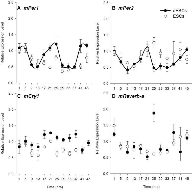
The appearance, progression, and potential role for circadian rhythms during early development have previously focused mainly on the suprachiasmatic nucleus (SCN) and peri- and postnatal expression of canonical clock genes. More recently, gene expression studies in embryonic stem cells have shown that some clock genes are expressed in undifferentiated cells; however rhythmicity was only established when cells are directed toward a neural fate. These studies also concluded that a functional clock is not present in ESCs, based solely on their gene expression. The null hypothesis underlying the present study is that embryonic stem cells become rhythmic in both clock gene expression and glucose utilization only when allowed to spontaneously differentiate. Undifferentiated stem cells (ESCs, n = 6 cultures/timepoint for all experiments) were either maintained in their pluripotent state or released into differentiation (dESCs, n = 6 cultures/timepoint for all experiments). Glucose utilization was assayed through 2-deoxyglucose uptake measurement, and clock gene and glucose transporter expression was assayed every 4 hours for 2 days in ESCs and dESCs by quantitative PCR (qPCR) in the same cell lysates. Undifferentiated stem cells expressed a self-sustained rhythm in glucose uptake that was not coincident with rhythmic expression of clock genes. This physiological rhythm was paralleled by glucose transporter mRNA expression. Upon differentiation, circadian patterns of some but not all clock genes were expressed, and the amplitude of the glucose utilization rhythm was enhanced in dESCs. These data provide the earliest evidence of a functional circadian clock, in addition to further challenging the idea that rhythmic transcription of clock genes are necessary for rhythmic physiological output and suggest a role for a clock-controlled physiology in the earliest stages of development.
昼夜节律在早期发育过程中的出现、进展和潜在作用以前主要集中在视交叉上核(SCN)和围产期及产后表达的经典时钟基因上。最近,胚胎干细胞中的基因表达研究表明,一些时钟基因在未分化细胞中表达;然而,只有当细胞向神经命运分化时,才会建立节律性。这些研究还得出结论,仅基于时钟基因的表达,胚胎干细胞中不存在功能性时钟。本研究的假设前提是,只有在允许胚胎干细胞自发分化的情况下,其时钟基因表达和葡萄糖利用才会呈现节律性。未分化的干细胞(ESCs,所有实验的每个时间点都有 6 个培养物)要么保持其多能状态,要么释放到分化中(dESCs,所有实验的每个时间点都有 6 个培养物)。葡萄糖利用通过 2-脱氧葡萄糖摄取测量来检测,时钟基因和葡萄糖转运蛋白的表达通过 qPCR 在相同的细胞裂解物中每隔 4 小时检测 2 天,在 ESCs 和 dESCs 中进行。未分化的干细胞表达出一种自我维持的葡萄糖摄取节律,与时钟基因的节律性表达不一致。这种生理节律与葡萄糖转运体 mRNA 的表达平行。分化后,一些(但不是所有)时钟基因的表达呈现出昼夜节律模式,并且 dESCs 中葡萄糖利用节律的幅度增强。这些数据提供了除进一步挑战时钟基因的节律性转录对于节律性生理输出是必要的这一观点之外,还提供了功能昼夜节律钟的最早证据,并表明时钟控制的生理学在发育的最早阶段发挥作用。