Cognitive Neuroscience Group, Department of Psychology, University of Amsterdam Weesperplein 4, 1018 XA, Amsterdam, The Netherlands ; Cognitive Science Center, University of Amsterdam Sarphatistraat 104, 1018 GV, Amsterdam, The Netherlands.
Brain Behav. 2012 Nov;2(6):763-77. doi: 10.1002/brb3.91. Epub 2012 Sep 29.
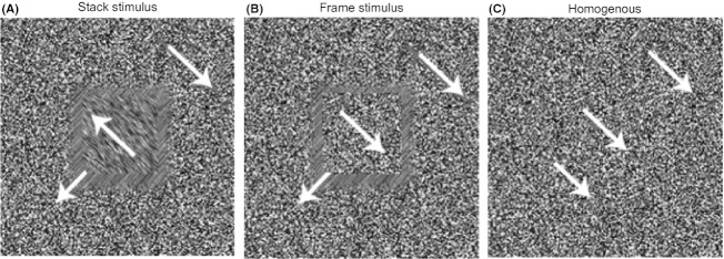
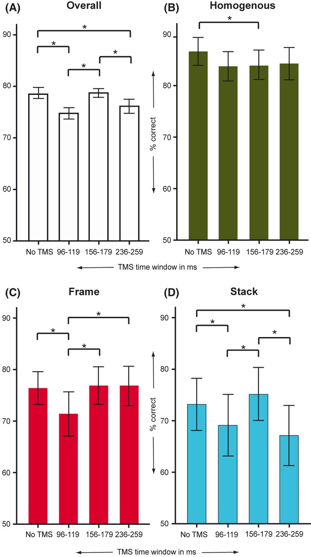
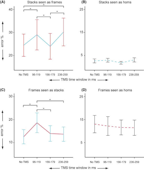
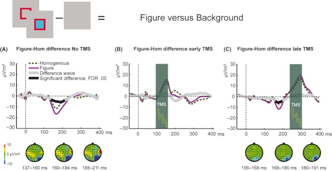
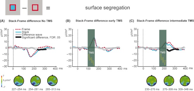
The ability to distinguish a figure from its background is crucial for visual perception. To date, it remains unresolved where and how in the visual system different stages of figure-ground segregation emerge. Neural correlates of figure border detection have consistently been found in early visual cortex (V1/V2). However, areas V1/V2 have also been frequently associated with later stages of figure-ground segregation (such as border ownership or surface segregation). To causally link activity in early visual cortex to different stages of figure-ground segregation, we briefly disrupted activity in areas V1/V2 at various moments in time using transcranial magnetic stimulation (TMS). Prior to stimulation we presented stimuli that made it possible to differentiate between figure border detection and surface segregation. We concurrently recorded electroencephalographic (EEG) signals to examine how neural correlates of figure-ground segregation were affected by TMS. Results show that disruption of V1/V2 in an early time window (96-119 msec) affected detection of figure stimuli and affected neural correlates of figure border detection, border ownership, and surface segregation. TMS applied in a relatively late time window (236-259 msec) selectively deteriorated performance associated with surface segregation. We conclude that areas V1/V2 are not only essential in an early stage of figure-ground segregation when figure borders are detected, but subsequently causally contribute to more sophisticated stages of figure-ground segregation such as surface segregation.
从背景中区分出一个图形对于视觉感知至关重要。迄今为止,视觉系统中不同的图形-背景分离阶段是在何处以及如何出现的,仍然没有得到解决。在早期视觉皮层(V1/V2)中一直发现有与图形边界检测相关的神经相关性。然而,V1/V2 区域也经常与图形-背景分离的后期阶段(例如边界所有权或表面分离)相关联。为了将早期视觉皮层的活动与图形-背景分离的不同阶段因果联系起来,我们使用经颅磁刺激(TMS)在不同时间点短暂地干扰 V1/V2 区域的活动。在刺激之前,我们呈现了可以区分图形边界检测和表面分离的刺激。我们同时记录脑电图(EEG)信号,以检查图形-背景分离的神经相关性如何受到 TMS 的影响。结果表明,V1/V2 在早期时间窗口(96-119 毫秒)中的干扰会影响图形刺激的检测,并影响图形边界检测、边界所有权和表面分离的神经相关性。在相对较晚的时间窗口(236-259 毫秒)应用 TMS 会选择性地降低与表面分离相关的性能。我们的结论是,V1/V2 区域不仅在检测图形边界时是图形-背景分离的早期阶段所必需的,而且随后还会对更复杂的图形-背景分离阶段(例如表面分离)产生因果影响。