Department of Ophthalmology, Vision Institute, UNIFESP, Hospital São Paulo-Federal University of São Paulo, São Paulo, São Paulo, Brazil.
BMJ Open. 2012 Nov 27;2(6). doi: 10.1136/bmjopen-2012-001266. Print 2012.
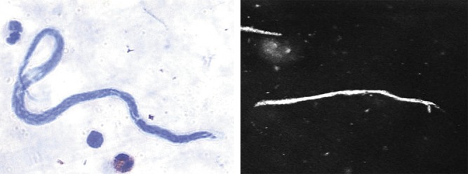
To characterise and confirm the presence of Mansonella ozzardi microfilariae in the cornea by biomicroscopy and corneal confocal microscopy.
Cross-sectional study.
Clinical practice study in patients from rural communities in Coari city on the Solimões river, Amazonas state, Brazil.
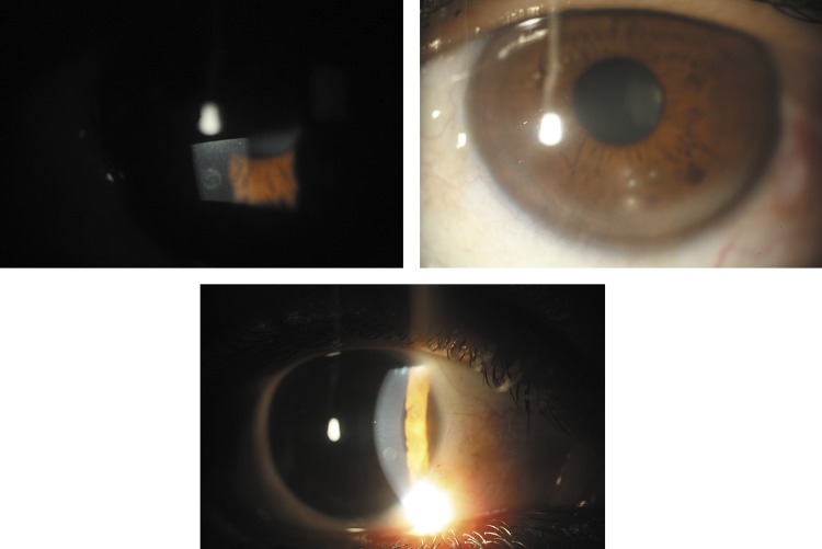
The eyes of 212 consecutive volunteer patients were examined using a flash light and their blood checked for the presence of microfilariae by an expert microscopist. Patients with suspicious corneal lesions (characterised as nummular keratitis) were submitted to biomicroscopy, fundoscopy and corneal confocal microscopy evaluation (CCME). In two patients, a biopsy of the limbal conjunctiva adjacent to the nummular keratitis was carried out and blood collected from the surgical wound for microfilariae investigation by thick blood film examination.
Positive correlation between corneal biomicroscopic and confocal lesions and M ozzardi microfilaremia.
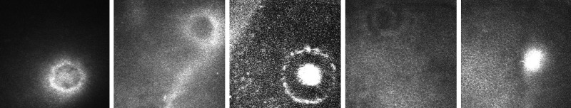
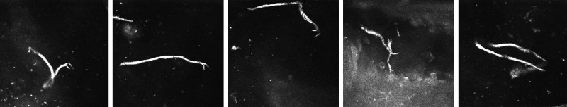
Of the 212 patients, 56 (26.4%) were positive for microfilaremia. 22 patients with nummular keratitis identified under flash light examination underwent biomicroscopy and CCME. Corneal lesions were positively correlated to microfilaremia (p=0.0001). At biomicroscopy, lesions were classified as quiescent or active. At CCME, lesions were categorised as circular or filiform. The associations between corneal lesions, CCME findings and microfilaremia are shown.
We describe M ozzardi microfilariae in the cornea and the associated eye pathology. Further studies using ocular tissue PCR and other imaging techniques would be helpful.
通过生物显微镜和角膜共聚焦显微镜来描述和确认曼森线虫微丝蚴在角膜中的存在。
横断面研究。
巴西亚马逊州索利蒙伊斯河畔科阿里市农村社区的临床实践研究。
对 212 名连续志愿患者的眼睛进行了检查,使用闪光灯,并由专家显微镜检查血液中是否存在微丝蚴。对疑似角膜病变(表现为钱币状角膜炎)的患者进行生物显微镜、眼底检查和角膜共聚焦显微镜评估(CCME)。在两名患者中,对紧邻钱币状角膜炎的角膜缘结膜进行了活检,并从手术伤口采集血液,通过厚血膜检查进行微丝蚴调查。
角膜生物显微镜和共聚焦病变与 M 奥兹ardi 微丝蚴血症之间的正相关。
在 212 名患者中,56 名(26.4%)微丝蚴血症阳性。在闪光灯检查下发现 22 名钱币状角膜炎患者进行了生物显微镜和 CCME 检查。角膜病变与微丝蚴血症呈正相关(p=0.0001)。在生物显微镜下,病变分为静止期或活动期。在 CCME 中,病变分为圆形或线状。显示了角膜病变、CCME 发现与微丝蚴血症之间的关联。
我们描述了曼森线虫微丝蚴在角膜中的存在及其相关的眼部病变。使用眼部组织 PCR 和其他成像技术进行进一步研究将有所帮助。