Rezaei-Kanavi Mozhgan, Javadi Mohammad-Ali, Mirbabaei-Ghafghazi Firooz
Eye Bank of I.R. Iran, Tehran, Iran.
J Ophthalmic Vis Res. 2009 Apr;4(2):122-4.
To describe the clinical and pathological features of a case of hydrogel intraocular lens (IOL) calcification.
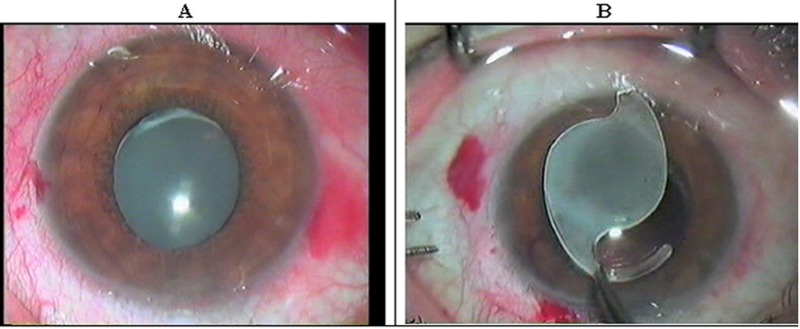
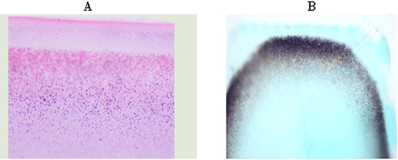
A 48-year-old man underwent explantation of a single-piece hydrophilic acrylic intraocular lens in his left eye because of decreased visual acuity and milky white opalescence of the IOL. The opacified lens was exchanged uneventfully with a hydrophobic acrylic IOL. Gross examination of the explanted IOL disclosed opacification of the optic and haptics. Full-thickness sections of the lens optic were stained with hematoxylin and eosin (H&E), von Kossa and Gram Tworts'. Microscopic examination of the sections revealed fine and diffuse basophilic granular deposits of variable size within the lens optic parallel to the lens curvature but separated from the surface by a moderately clear zone. The deposits were of high calcium content as evident by dark brown staining with von Kossa. Gram Tworts' staining disclosed no microorganisms.
This report further contributes to the existing literature on hydrogel IOL calcification.
描述一例水凝胶人工晶状体(IOL)钙化的临床和病理特征。
一名48岁男性因视力下降和IOL呈乳白色浑浊,接受了左眼单片亲水性丙烯酸人工晶状体的取出术。浑浊的晶状体顺利更换为疏水性丙烯酸IOL。对取出的IOL进行大体检查发现光学部和襻部浑浊。晶状体光学部的全层切片用苏木精和伊红(H&E)、冯·科萨染色法和革兰染色法染色。对切片进行显微镜检查发现,在晶状体光学部内有大小不一的细小弥漫性嗜碱性颗粒沉积物,与晶状体曲率平行,但与表面之间有一个中度清晰的区域。通过冯·科萨染色呈深棕色可见,这些沉积物钙含量很高。革兰染色未发现微生物。
本报告进一步丰富了关于水凝胶IOL钙化的现有文献。