• 文献检索
  • 文档翻译
  • 深度研究
  • 学术资讯
  • Suppr Zotero 插件Zotero 插件
  • 邀请有礼
  • 套餐&价格
  • 历史记录
应用&插件
Suppr Zotero 插件Zotero 插件浏览器插件Mac 客户端Windows 客户端微信小程序
定价
高级版会员购买积分包购买API积分包
服务
文献检索文档翻译深度研究API 文档MCP 服务
关于我们
关于 Suppr公司介绍联系我们用户协议隐私条款
关注我们

Suppr 超能文献

核心技术专利:CN118964589B侵权必究
粤ICP备2023148730 号-1Suppr @ 2026

文献检索

告别复杂PubMed语法,用中文像聊天一样搜索,搜遍4000万医学文献。AI智能推荐,让科研检索更轻松。

立即免费搜索

文件翻译

保留排版,准确专业,支持PDF/Word/PPT等文件格式,支持 12+语言互译。

免费翻译文档

深度研究

AI帮你快速写综述,25分钟生成高质量综述,智能提取关键信息,辅助科研写作。

立即免费体验

后弹力层剥除内皮角膜移植术后亲水性人工晶状体表面沉积物的物理化学分析

Physicochemical Analysis of Sediments Formed on the Surface of Hydrophilic Intraocular Lens after Descemet's Stripping Endothelial Keratoplasty.

作者信息

Tarnawska Dorota, Balin Katarzyna, Jastrzębska Maria, Talik Agnieszka, Wrzalik Roman

机构信息

Institute of Biomedical Engineering, Faculty of Science and Technology, University of Silesia, 41-200 Sosnowiec, Poland.

Department of Ophthalmology, District Railway Hospital, Panewnicka 65, 40-760 Katowice, Poland.

出版信息

Materials (Basel). 2020 Sep 17;13(18):4145. doi: 10.3390/ma13184145.

DOI:10.3390/ma13184145
PMID:32957729
原文链接:https://pmc.ncbi.nlm.nih.gov/articles/PMC7560278/
Abstract

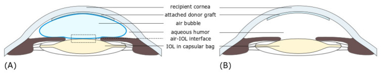
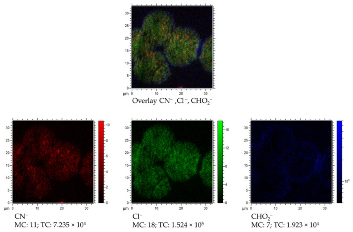
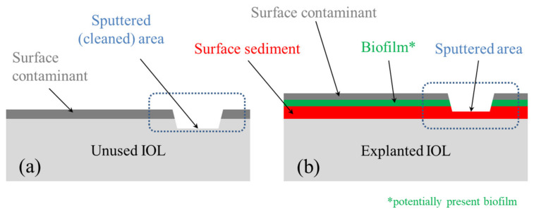
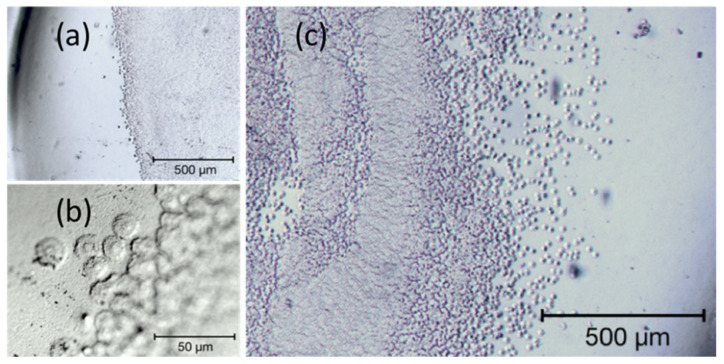
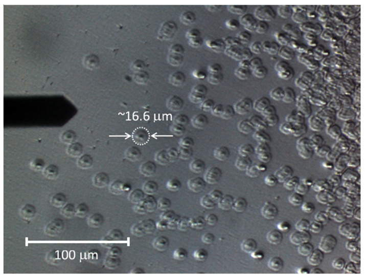
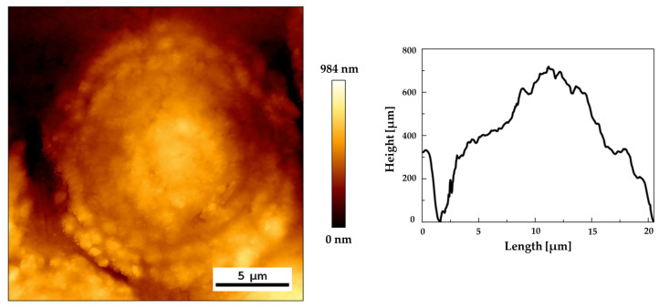
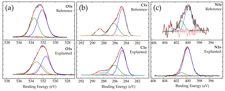
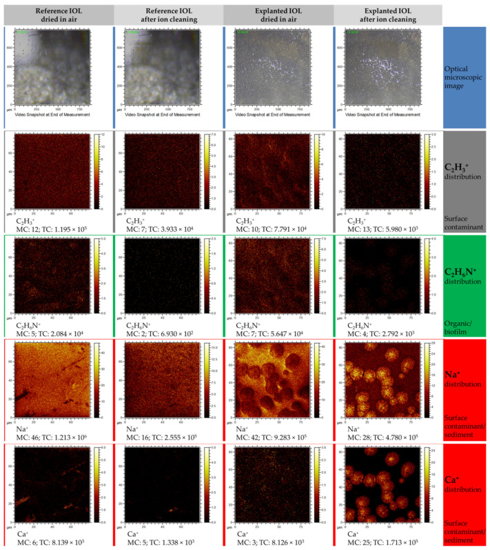
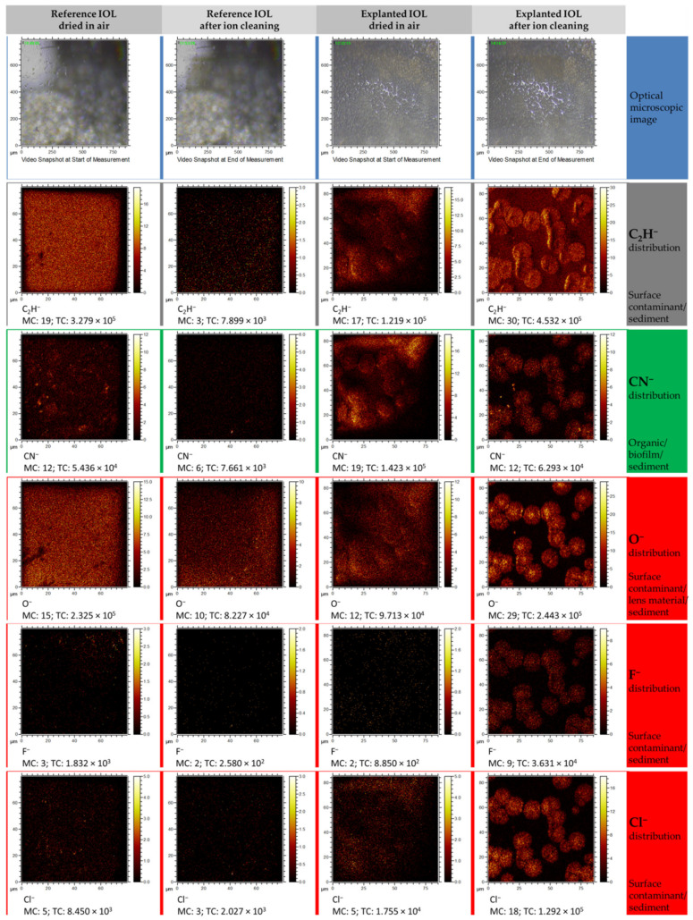
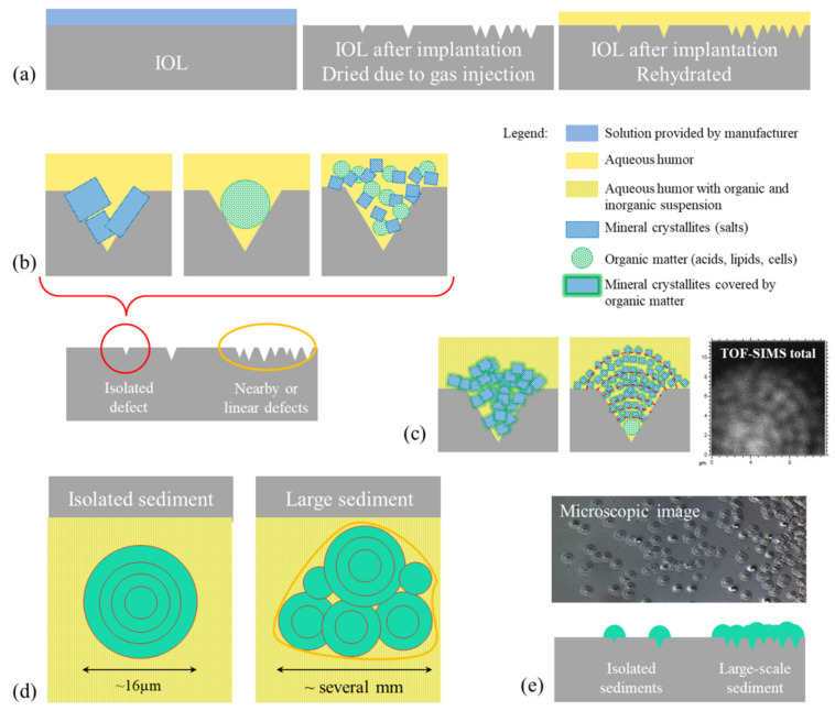
An intraocular lens (IOL) is a synthetic, artificial lens placed inside the eye that replaces a natural lens that is surgically removed, usually as part of cataract surgery. The opacification of the artificial lens can be related to the formation of the sediments on its surface and could seriously impair vision. The physicochemical analysis was performed on an explanted hydrophilic IOL and compared to the unused one, considered as a reference IOL. The studies were carried out using surface sensitive techniques, which can contribute to a better understanding of the sedimentation process on hydrophilic IOLs' surfaces. The microscopic studies allowed us to determine the morphology of sediments observed on explanted IOL. The photoelectron spectroscopy measurements revealed the presence of organic and inorganic compounds at the lens surface. Mass spectroscopy measurements confirmed the chemical composition of deposits and allowed for chemical imaging of the IOL surface. Applied techniques allowed to obtain a new set of information approximating the origin of the sediments' formation on the surface of the hydrophilic IOLs after Descemet's stripping endothelial keratoplasty.

摘要

人工晶状体(IOL)是一种放置在眼内的合成人造晶状体,用于替代通常在白内障手术中被手术摘除的天然晶状体。人工晶状体的混浊可能与其表面沉积物的形成有关,并可能严重损害视力。对一个摘除的亲水性人工晶状体进行了物理化学分析,并与未使用的人工晶状体(作为参考人工晶状体)进行了比较。研究采用了表面敏感技术,这有助于更好地理解亲水性人工晶状体表面的沉积过程。显微镜研究使我们能够确定在摘除的人工晶状体上观察到的沉积物的形态。光电子能谱测量揭示了晶状体表面存在有机和无机化合物。质谱测量证实了沉积物的化学成分,并实现了人工晶状体表面的化学成像。所应用的技术使我们能够获得一组新的信息,这些信息近似于在Descemet膜剥脱性内皮角膜移植术后亲水性人工晶状体表面沉积物形成的起源。

https://cdn.ncbi.nlm.nih.gov/pmc/blobs/5a73/7560278/6c0577673a78/materials-13-04145-g010.jpg
https://cdn.ncbi.nlm.nih.gov/pmc/blobs/5a73/7560278/06a116d16285/materials-13-04145-g001.jpg
https://cdn.ncbi.nlm.nih.gov/pmc/blobs/5a73/7560278/66d86bd4b80a/materials-13-04145-g002.jpg
https://cdn.ncbi.nlm.nih.gov/pmc/blobs/5a73/7560278/ee0d374ee786/materials-13-04145-g003.jpg
https://cdn.ncbi.nlm.nih.gov/pmc/blobs/5a73/7560278/e4e704ed1299/materials-13-04145-g004.jpg
https://cdn.ncbi.nlm.nih.gov/pmc/blobs/5a73/7560278/ca716af7df5b/materials-13-04145-g005.jpg
https://cdn.ncbi.nlm.nih.gov/pmc/blobs/5a73/7560278/973ebcb73db7/materials-13-04145-g006.jpg
https://cdn.ncbi.nlm.nih.gov/pmc/blobs/5a73/7560278/1844aa50e768/materials-13-04145-g007.jpg
https://cdn.ncbi.nlm.nih.gov/pmc/blobs/5a73/7560278/ca00c92b7424/materials-13-04145-g008.jpg
https://cdn.ncbi.nlm.nih.gov/pmc/blobs/5a73/7560278/516867afc728/materials-13-04145-g009.jpg
https://cdn.ncbi.nlm.nih.gov/pmc/blobs/5a73/7560278/6c0577673a78/materials-13-04145-g010.jpg

相似文献

1
Physicochemical Analysis of Sediments Formed on the Surface of Hydrophilic Intraocular Lens after Descemet's Stripping Endothelial Keratoplasty.后弹力层剥除内皮角膜移植术后亲水性人工晶状体表面沉积物的物理化学分析
Materials (Basel). 2020 Sep 17;13(18):4145. doi: 10.3390/ma13184145.
2
Intraocular lens opacification after Descemet's stripping automated endothelial keratoplasty.Descemet膜剥除自动内皮角膜移植术后人工晶状体混浊
Taiwan J Ophthalmol. 2017 Jul-Sep;7(3):160-163. doi: 10.4103/tjo.tjo_54_17.
3
Anterior surface opacification of intraocular lenses after Descemet's stripping automated endothelial keratoplasty.后弹力层剥除自动角膜内皮移植术后人工晶状体前表面混浊
BMJ Case Rep. 2015 Dec 16;2015:bcr2015213216. doi: 10.1136/bcr-2015-213216.
4
[Intraocular Lens Opacification Following Posterior Lamellar Keratoplasty].[后板层角膜移植术后人工晶状体混浊]
Klin Monbl Augenheilkd. 2015 Aug;232(8):976-81. doi: 10.1055/s-0035-1546175. Epub 2015 Aug 19.
5
[Opacification of a hydrophilic acrylic intraocular lens after DMEK : A material analysis].[DMEK术后亲水性丙烯酸人工晶状体混浊:材料分析]
Ophthalmologe. 2017 Sep;114(9):832-837. doi: 10.1007/s00347-016-0394-6.
6
Hydrophilic intraocular lens opacification after posterior lamellar keratoplasty - a material analysis with special reference to optical quality assessment.后板层角膜移植术后亲水性人工晶状体混浊——特别涉及光学质量评估的材料分析
BMC Ophthalmol. 2017 Aug 22;17(1):150. doi: 10.1186/s12886-017-0546-8.
7
Intraocular lens opacification after corneal endothelial keratoplasty: electron microscopy and x-ray element spectroscopy analysis.角膜内皮移植术后人工晶状体混浊:电子显微镜及X射线元素光谱分析
J Cataract Refract Surg. 2015 Jan;41(1):140-5. doi: 10.1016/j.jcrs.2014.05.035.
8
Hydrophilic Acrylic Intraocular Lens Opacification after Descemet Stripping Automated Endothelial Keratoplasty.深板层角膜内皮移植术后亲水性丙烯酸人工晶状体混浊
J Ophthalmic Vis Res. 2016 Apr-Jun;11(2):225-7. doi: 10.4103/2008-322X.158897.
9
Opacification of a hydrophilic acrylic intraocular lens with a hydrophobic surface after air injection in Descemet-stripping automated endothelial keratoplasty in a patient with Fuchs dystrophy.在一名患有富克斯角膜内皮营养不良的患者接受后弹力层剥除自动角膜内皮移植术时,向眼内注入空气后,表面疏水的亲水性丙烯酸人工晶状体发生了混浊。
J Cataract Refract Surg. 2016 Mar;42(3):485-8. doi: 10.1016/j.jcrs.2016.02.004. Epub 2016 Mar 19.
10
[Post operation complications analysis and their treatment of Descemet's stripping automated endothelium keratoplasty].[后弹力层剥除自动角膜内皮移植术的术后并发症分析及其治疗]
Zhonghua Yan Ke Za Zhi. 2014 Apr;50(4):254-60.

引用本文的文献

1
Impact of Calcium and Phosphorus Levels on Optical Deterioration in Primary and Secondary Intraocular Lens Calcification.钙磷水平对原发性和继发性眼内晶状体钙化光学恶化的影响。
Transl Vis Sci Technol. 2024 Oct 1;13(10):18. doi: 10.1167/tvst.13.10.18.
2
Current State of the Art and Next Generation of Materials for a Customized IntraOcular Lens according to a Patient-Specific Eye Power.根据患者特定眼屈光度定制人工晶状体的当前技术水平及下一代材料
Polymers (Basel). 2023 Mar 22;15(6):1590. doi: 10.3390/polym15061590.
3
Development of a standardized in vitro model to reproduce hydrophilic acrylic intraocular lens calcification.

本文引用的文献

1
Serial opacification of a hydrophilic-hydrophobic acrylic intraocular lens: analysis of potential risk factors.亲水性-疏水性丙烯酸眼内晶状体的连续混浊:潜在危险因素分析。
J Cataract Refract Surg. 2020 Dec;46(12):1624-1629. doi: 10.1097/j.jcrs.0000000000000342.
2
Variation in intraocular lens calcification under different environmental conditions in eyes with supplementary sulcus-supported lenses.在植入有辅助性沟支撑人工晶状体的眼中,不同环境条件下人工晶状体钙化的差异。
Am J Ophthalmol Case Rep. 2020 Jun 24;19:100797. doi: 10.1016/j.ajoc.2020.100797. eCollection 2020 Sep.
3
Reasons for explantation, demographics, and material analysis of 200 intraocular lens explants.
开发一种标准化的体外模型以重现亲水性丙烯酸人工晶状体钙化。
Sci Rep. 2022 May 10;12(1):7685. doi: 10.1038/s41598-022-11486-0.
4
The Structure-Properties-Cytotoxicity Interplay: A Crucial Pathway to Determining Graphene Oxide Biocompatibility.结构-性质-细胞毒性相互作用:决定氧化石墨烯生物相容性的关键途径。
Int J Mol Sci. 2021 May 20;22(10):5401. doi: 10.3390/ijms22105401.
5
Analysis of opacification patterns in intraocular lenses (IOL).人工晶状体(IOL)混浊模式分析。
BMJ Open Ophthalmol. 2021 Feb 11;6(1):e000589. doi: 10.1136/bmjophth-2020-000589. eCollection 2021.
200 枚人工晶状体取出的原因、人口统计学资料和材料分析。
J Cataract Refract Surg. 2020 Jan;46(1):20-26. doi: 10.1016/j.jcrs.2019.08.045.
4
Localized calcification of hydrophilic acrylic intraocular lenses after posterior segment procedures.后段手术后继发性亲水丙烯酸人工晶状体局限性钙化。
J Cataract Refract Surg. 2019 Dec;45(12):1801-1807. doi: 10.1016/j.jcrs.2019.07.015.
5
Systematic review of potential causes of intraocular lens opacification.系统评价导致人工晶状体混浊的潜在原因。
Clin Exp Ophthalmol. 2020 Jan;48(1):89-97. doi: 10.1111/ceo.13650. Epub 2019 Oct 22.
6
Calcification of hydrophilic acrylic intraocular lenses following secondary surgical procedures in the anterior and posterior segments.眼后段和眼前段二次手术后继发亲水性丙烯酸人工晶状体钙化。
Br J Ophthalmol. 2019 Dec;103(12):1700-1703. doi: 10.1136/bjophthalmol-2018-313385. Epub 2019 Feb 23.
7
Large-scale opacification of a hydrophilic/hydrophobic intraocular lens.亲水性/疏水性人工晶状体的大规模浑浊。
Eur J Ophthalmol. 2020 Mar;30(2):307-314. doi: 10.1177/1120672119830581. Epub 2019 Feb 19.
8
Opacification of hydrophilic intraocular lenses associated with vitrectomy and injection of intraocular gas.亲水型人工晶状体混浊与玻璃体切除术及眼内气体注射相关。
BMJ Open Ophthalmol. 2018 Dec 15;3(1):e000157. doi: 10.1136/bmjophth-2018-000157. eCollection 2018.
9
[Opacification of hydrophilic intraocular lens after multiple injections of bevacizumab].[多次注射贝伐单抗后亲水人工晶状体混浊]
Ophthalmologe. 2019 Sep;116(9):882-886. doi: 10.1007/s00347-018-0829-3.
10
Serial intraocular lens opacifications of different designs from the same manufacturer: Clinical and light microscopic results of 71 explant cases.同一制造商不同设计的连续眼内晶状体混浊:71 例植入物病例的临床和光学显微镜结果。
J Cataract Refract Surg. 2018 Nov;44(11):1326-1332. doi: 10.1016/j.jcrs.2018.07.026. Epub 2018 Sep 29.