Max Planck Institute for Developmental Biology, Spemannstrasse 35, Tübingen, 72076, Germany.
Evodevo. 2012 Dec 3;3(1):27. doi: 10.1186/2041-9139-3-27.
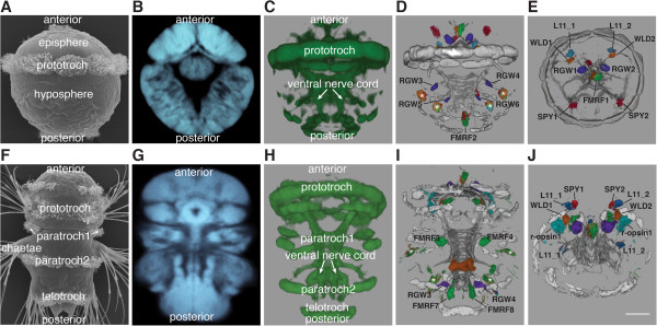
Digital anatomical atlases are increasingly used in order to depict different gene expression patterns and neuronal morphologies within a standardized reference template. In evo-devo, a discipline in which the comparison of gene expression patterns is a widely used approach, such standardized anatomical atlases would allow a more rigorous assessment of the conservation of and changes in gene expression patterns during micro- and macroevolutionary time scales. Due to its small size and invariant early development, the annelid Platynereis dumerilii is particularly well suited for such studies. Recently a reference template with registered gene expression patterns has been generated for the anterior part (episphere) of the Platynereis trochophore larva and used for the detailed study of neuronal development.
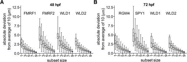
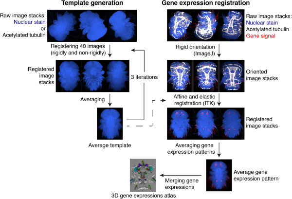
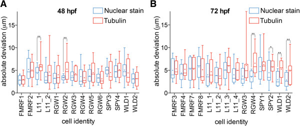
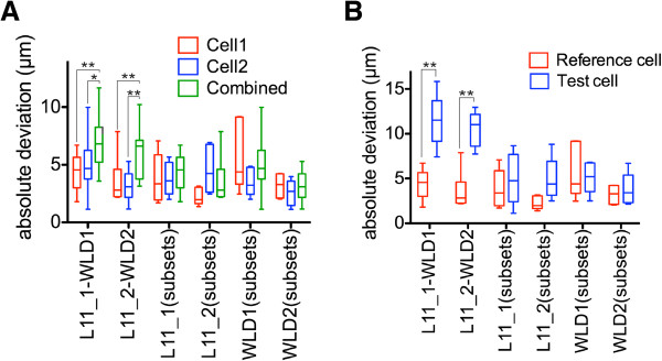
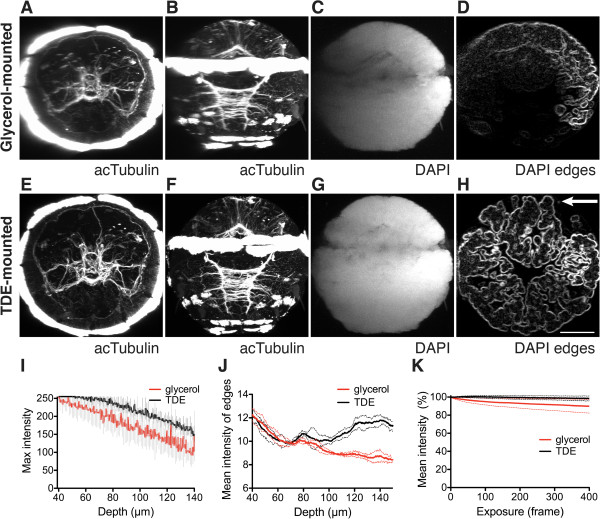
Here we introduce and evaluate a method for whole-body gene expression pattern registration for Platynereis trochophore and nectochaete larvae based on whole-mount in situ hybridization, confocal microscopy, and image registration. We achieved high-resolution whole-body scanning using the mounting medium 2,2'-thiodiethanol (TDE), which allows the matching of the refractive index of the sample to that of glass and immersion oil thereby reducing spherical aberration and improving depth penetration. This approach allowed us to scan entire whole-mount larvae stained with nitroblue tetrazolium/5-bromo-4-chloro-3-indolyl phosphate (NBT/BCIP) in situ hybridization and counterstained fluorescently with an acetylated-tubulin antibody and the nuclear stain 4'6-diamidino-2-phenylindole (DAPI). Due to the submicron isotropic voxel size whole-mount larvae could be scanned in any orientation. Based on the whole-body scans, we generated four different reference templates by the iterative registration and averaging of 40 individual image stacks using either the acetylated-tubulin or the nuclear-stain signal for each developmental stage. We then registered to these templates the expression patterns of cell-type specific genes. In order to evaluate the gene expression pattern registration, we analyzed the absolute deviation of cell-center positions. Both the acetylated-tubulin- and the nuclear-stain-based templates allowed near-cellular-resolution gene expression registration. Nuclear-stain-based templates often performed significantly better than acetylated-tubulin-based templates. We provide detailed guidelines and scripts for the use and further expansion of the Platynereis gene expression atlas.
We established whole-body reference templates for the generation of gene expression atlases for Platynereis trochophore and nectochaete larvae. We anticipate that nuclear-staining-based image registration will be applicable for whole-body alignment of the embryonic and larval stages of other organisms in a similar size range.
为了在标准化参考模板中描绘不同的基因表达模式和神经元形态,数字解剖图谱的使用越来越多。在进化发育生物学中,比较基因表达模式是一种广泛使用的方法,这种标准化的解剖图谱将允许更严格地评估在微观和宏观进化时间尺度上基因表达模式的保守性和变化。由于其体积小且早期发育不变,环节动物扁形虫 Platynereis dumerilii 特别适合此类研究。最近,已经为扁形虫担轮幼虫的前(赤道)部分生成了具有注册基因表达模式的参考模板,并用于详细研究神经元发育。
在这里,我们介绍并评估了一种基于全胚胎原位杂交、共聚焦显微镜和图像配准的扁形虫担轮幼虫和幼体整体基因表达模式配准方法。我们使用 mounting medium 2,2'-thiodiethanol (TDE) 实现了高分辨率的全身体扫描,该方法允许样品的折射率与玻璃和浸油相匹配,从而减少球差并提高深度穿透。这种方法使我们能够扫描整个用硝基蓝四唑/5-溴-4-氯-3-吲哚磷酸(NBT/BCIP)原位杂交染色并用抗乙酰化微管蛋白抗体和核染剂 4'6-二脒基-2-苯基吲哚(DAPI)荧光染色的全胚胎幼虫。由于亚微米各向同性体素大小,全胚胎幼虫可以以任何方向进行扫描。基于全身体扫描,我们通过迭代注册和平均 40 个个体图像堆栈生成了四个不同的参考模板,每个发育阶段都使用抗乙酰化微管蛋白或核染色信号。然后,我们将细胞类型特异性基因的表达模式注册到这些模板上。为了评估基因表达模式注册,我们分析了细胞中心位置的绝对偏差。抗乙酰化微管蛋白和核染色基础模板都允许接近细胞分辨率的基因表达注册。核染色基础模板的性能通常明显优于抗乙酰化微管蛋白基础模板。我们提供了使用和进一步扩展扁形虫基因表达图谱的详细指南和脚本。
我们为扁形虫担轮幼虫和幼体建立了整体参考模板,用于生成基因表达图谱。我们预计核染色的图像注册将适用于类似大小范围的其他生物体的胚胎和幼虫阶段的全身体对齐。