Department of Radiology, Renji Hospital, Shanghai Jiao Tong University School of Medicine, Shanghai, China.
PLoS One. 2012;7(11):e50319. doi: 10.1371/journal.pone.0050319. Epub 2012 Nov 28.
To characterize lymphatic vessel morphology in lower extremity lymphedema using MR lymphography at 3T.
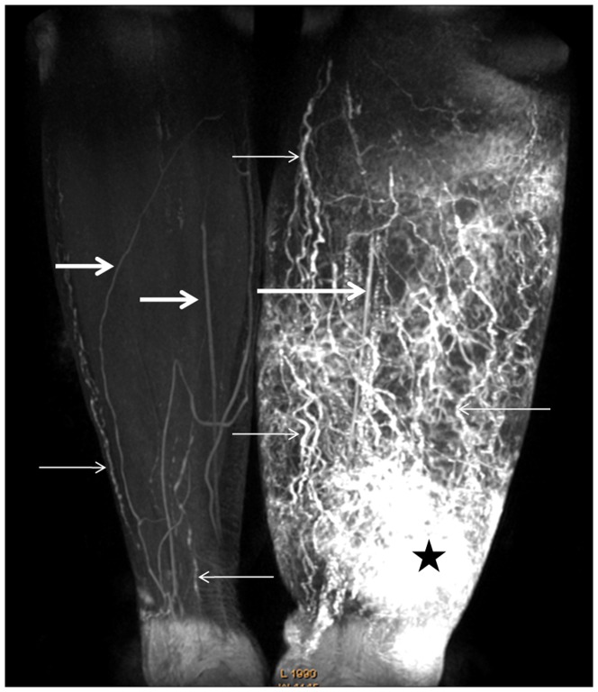
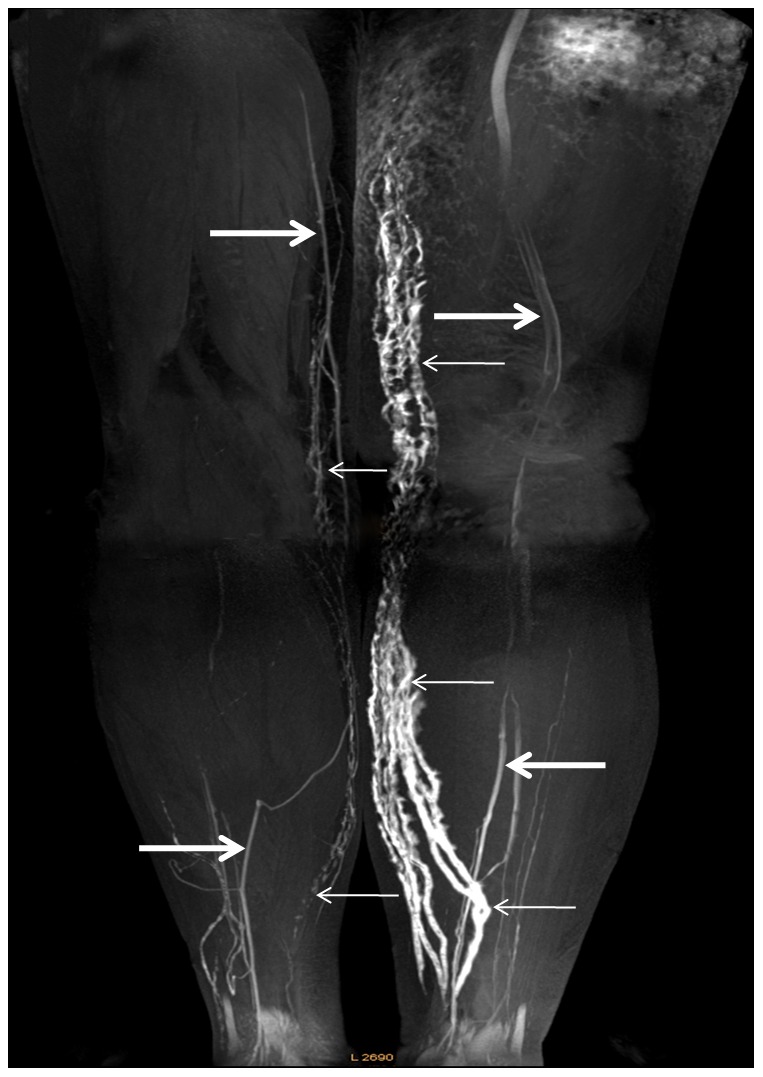
Forty females with lower extremity lymphedema secondary to gynecologic carcinoma treatment underwent MR lymphography (MRL) at 3T. Lymphatic vessel morphology in normal and affected limbs was compared.
The median diameter of the lymphatic vessels in swollen calf and thigh were significantly larger than that in the contralateral calf and thigh, respectively (p<0.05). The median number of lymphatic vessels visualized in normal calf was less than that in the lymphedematous calf (p<0.01), while no significant difference was found between the normal thigh and swollen thigh. Lymphatic vessel number in the affected calf was significantly greater than that in affected thigh and the mean diameter of affected calf was also significantly wider than that of affected thigh (p<0.01). Mean diameter of lymphatic vessels in the affected calf was significantly different between stage I and stage III (p<0.05), but not significantly different between stages I and II, and between stages II and III (p>0.05). The median number of lymphatic vessels for affected calf showed significant difference between stage I and stage III, and between stage II and stage III (p<0.05), but no significant difference between stage I and stage II (p>0.05). There was no significant difference in mean diameter or median number of lymphatic vessels in the affected thigh found between different stages (p>0.05).
There are significant differences in the number or diameter of lymphatic vessels between normal and affected limbs and there are significant differences for affected calf between early and late stages of lymphedema; therefore, MR lymphography can be helpful in diagnosis or clinical staging for lower extremity with gynecologic oncology-related lymphedema.
在 3T 磁共振淋巴造影术下对下肢淋巴水肿的淋巴管形态进行特征分析。
40 名女性因妇科癌症治疗而导致下肢淋巴水肿,在 3T 磁共振淋巴造影术下进行淋巴造影术检查。比较正常和患病肢体的淋巴管形态。
肿胀的小腿和大腿的淋巴管直径中位数明显大于对侧小腿和大腿(p<0.05)。正常小腿可见的淋巴管中位数少于淋巴水肿小腿(p<0.01),而正常大腿与肿胀大腿之间无显著差异。患病小腿的淋巴管数量明显大于患病大腿,且患病小腿的平均直径也明显大于患病大腿(p<0.01)。患病小腿的淋巴管直径在 1 期和 3 期之间存在显著差异(p<0.05),但在 1 期和 2 期之间以及 2 期和 3 期之间无显著差异(p>0.05)。患病小腿的淋巴管中位数在 1 期和 3 期之间、2 期和 3 期之间存在显著差异(p<0.05),但在 1 期和 2 期之间无显著差异(p>0.05)。患病大腿的淋巴管平均直径或中位数在不同阶段之间无显著差异(p>0.05)。
正常和患病肢体的淋巴管数量或直径存在显著差异,并且在淋巴水肿的早期和晚期,患病小腿之间存在显著差异;因此,磁共振淋巴造影术有助于诊断或临床分期妇科肿瘤相关的下肢淋巴水肿。