Astvaldsdóttir A, Ahlund K, Holbrook W P, de Verdier B, Tranæus S
Division of Cariology, Department of Dental Medicine, Karolinska Institutet, Huddinge, Sweden ; Faculty of Odontology, University of Iceland, Reykjavík, Iceland.
Int J Dent. 2012;2012:326401. doi: 10.1155/2012/326401. Epub 2012 Nov 4.
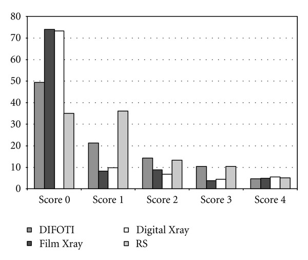
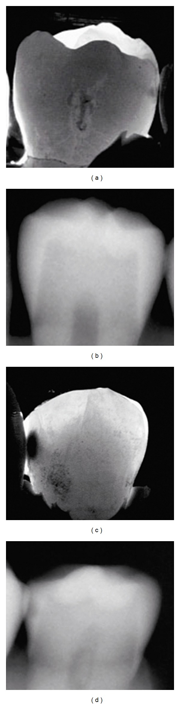
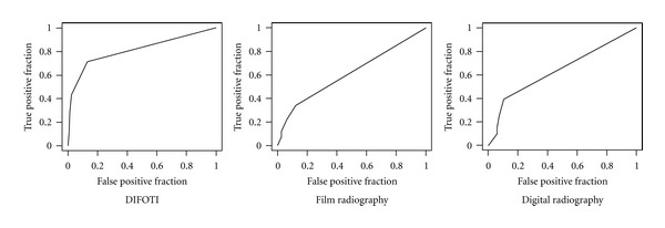
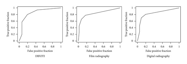
The aim of the present study was to compare the diagnostic accuracy/efficacy of digital imaging fiber-optic transillumination (DIFOTI) with film and digital radiography, in detection of approximal caries lesions. One hundred and twelve approximal surfaces were scored for caries, using DIFOTI images film and digital radiographs. All three sets of images were examined twice by 8 observers, with a minimal interval of one week between examinations. Validation of histological sections served as a reference standard. Reproducibility, based on intra- and interobserver agreement, was similar for all three methods. At diagnostic threshold D1 (enamel and dentin caries), DIFOTI showed significantly higher sensitivity, but differences in specificity between methods were nonsignificant. Diagnostic accuracy in the form of area under the receiver operating characteristic curve (AUC) was significantly higher for DIFOTI. At diagnostic threshold D3 (dentin caries), the differences in sensitivity and AUC among methods were nonsignificant, but DIFOTI showed significantly lower specificity. Compared with the radiographs, DIFOTI showed closer agreement, expressed as weighted kappa values, with the reference standard. The results show that under in vitro conditions, the diagnostic accuracy of DIFOTI in detecting early approximal enamel lesions is greater than that of film and digital radiography, while the potential for detecting lesions in dentin is similar for all three methods.
本研究的目的是比较数字成像光纤透照术(DIFOTI)与胶片及数字X线摄影在检测邻面龋损方面的诊断准确性/效能。使用DIFOTI图像、胶片和数字X线照片对112个邻面进行龋损评分。8名观察者对所有三组图像进行两次检查,两次检查之间的间隔至少为一周。组织学切片验证作为参考标准。基于观察者内和观察者间一致性的可重复性,三种方法相似。在诊断阈值D1(釉质和牙本质龋)时,DIFOTI显示出显著更高的敏感性,但方法间特异性差异不显著。以受试者工作特征曲线下面积(AUC)形式表示的诊断准确性,DIFOTI显著更高。在诊断阈值D3(牙本质龋)时,方法间敏感性和AUC的差异不显著,但DIFOTI显示出显著更低的特异性。与X线照片相比,DIFOTI与参考标准的一致性更紧密,以加权kappa值表示。结果表明,在体外条件下,DIFOTI在检测早期邻面釉质病变方面的诊断准确性高于胶片和数字X线摄影,而三种方法检测牙本质病变的潜力相似。