Greenberg Division of Cardiology, Weill Cornell Medical College , New York, NY 10065 , USA.
Biol Open. 2012 Mar 15;1(3):208-19. doi: 10.1242/bio.2012038. Epub 2012 Jan 19.
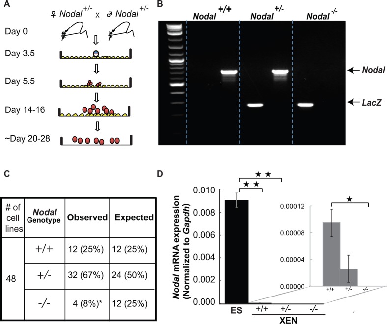
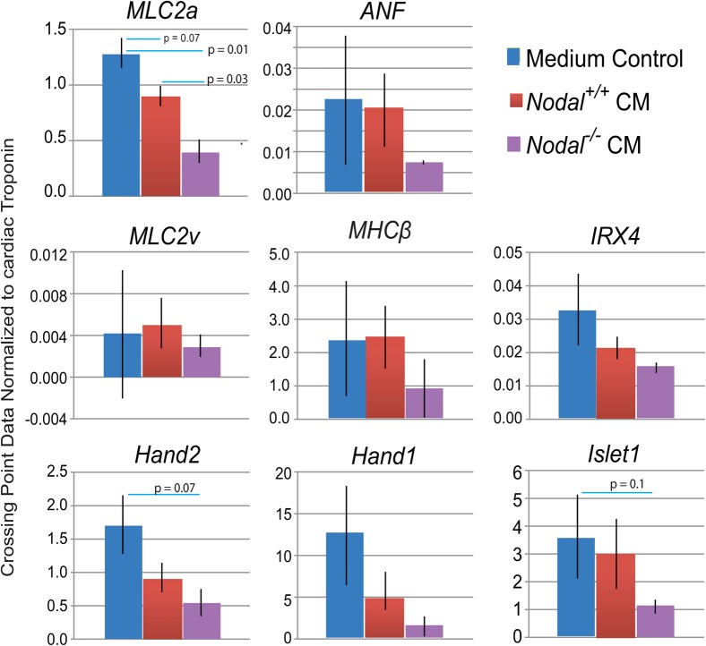
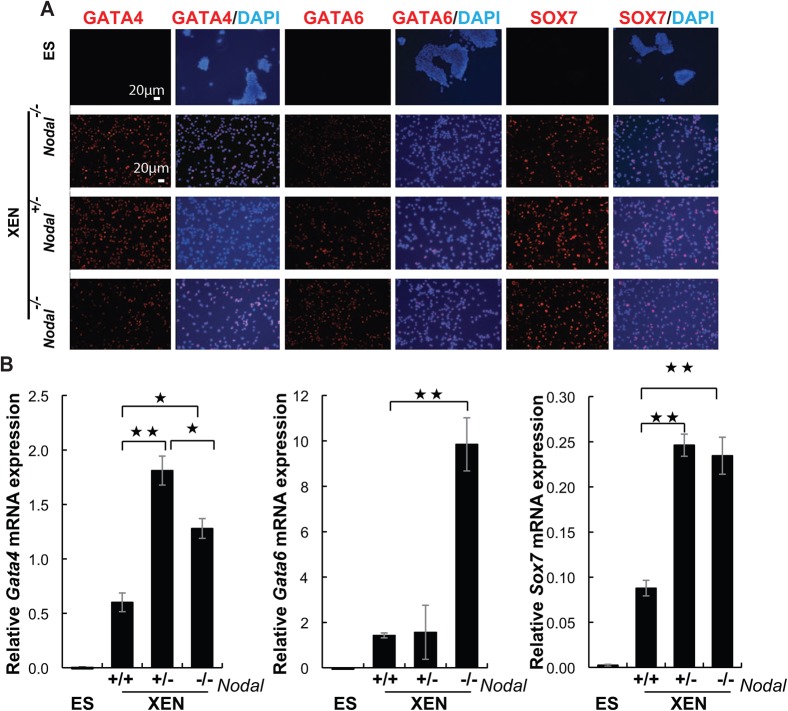
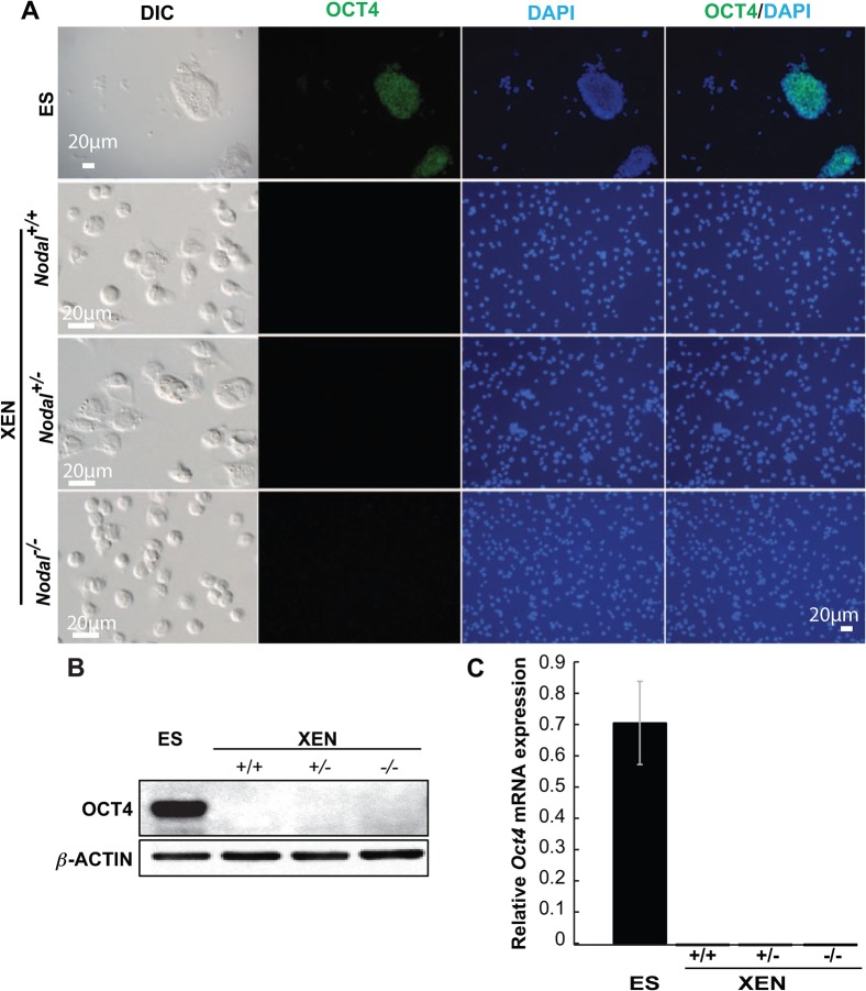
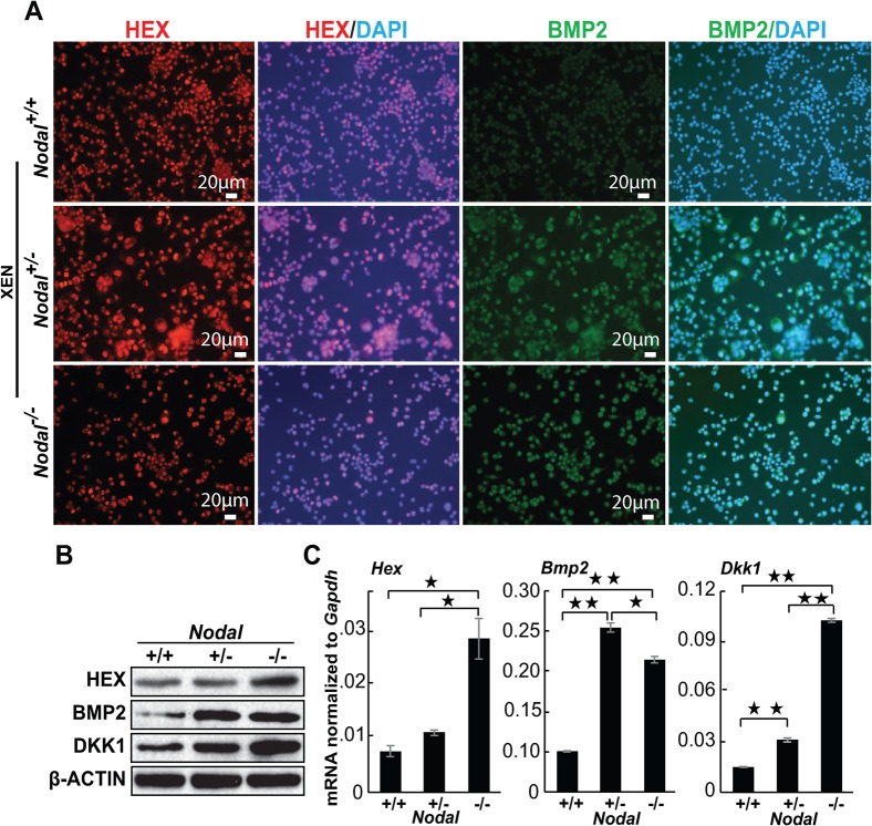
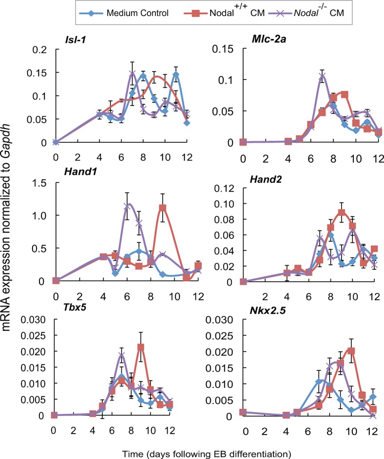
Interactions between the endoderm and mesoderm that mediate myocardial induction are difficult to study in vivo because of the small size of mammalian embryos at relevant stages. However, we and others have demonstrated that signals from endodermal cell lines can influence myocardial differentiation from both mouse and human embryoid bodies (EBs), and because of this, assays that utilize embryonic stem (ES) cells and endodermal cell lines provide excellent in vitro models to study early cardiac differentiation. Extraembryonic endoderm (XEN) stem cells have a particular advantage over other heart-inducing cell lines in that they can easily be derived from both wild type and mutant mouse blastocysts. Here we describe the first isolation of a Nodal mutant XEN stem cell line. Nodal(-/-) XEN cell lines were not isolated at expected Mendelian ratios, and those that were successfully established, showed an increase in markers for the anterior visceral endoderm (AVE). Since AVE represents the heart-inducing endoderm in the mouse, cardiac differentiation was compared in EBs treated with conditioned medium (CM) collected from wild type or Nodal(-/-) XEN cells. EBs treated with CM from Nodal(-/-) cells began beating earlier and showed early activation of myocardial genes, but this early cardiac differentiation did not cause an overall increase in cardiomyocyte yield. By comparison, CM from wild type XEN cells both delayed cardiac differentiation and caused a concomitant increase in overall cardiomyocyte formation. Detailed marker analysis suggested that early activation of cardiac differentiation by Nodal(-/-) XEN CM caused premature differentiation and subsequent depletion of cardiac progenitors.
内胚层和中胚层之间介导心肌诱导的相互作用在体内很难研究,因为在相关阶段哺乳动物胚胎的体积很小。然而,我们和其他人已经证明,来自内胚层细胞系的信号可以影响来自小鼠和人类胚状体 (EB) 的心肌分化,因此,利用胚胎干细胞 (ES) 细胞和内胚层细胞系的测定提供了极好的体外模型来研究早期心脏分化。胚胎外内胚层 (XEN) 干细胞在诱导心脏方面比其他诱导心脏的细胞系具有特殊的优势,因为它们可以很容易地从野生型和突变型小鼠胚泡中分离出来。在这里,我们描述了第一个 Nodal 突变 XEN 干细胞系的分离。Nodal(-/-) XEN 细胞系没有按照预期的孟德尔比例分离出来,那些成功建立的细胞系显示出前内脏内胚层 (AVE) 的标志物增加。由于 AVE 代表了小鼠中的心脏诱导内胚层,因此比较了用来自野生型或 Nodal(-/-) XEN 细胞的条件培养基 (CM) 处理的 EB 中的心脏分化。用 Nodal(-/-) 细胞的 CM 处理的 EB 更早开始跳动,并显示出心肌基因的早期激活,但这种早期心脏分化并没有导致心肌细胞产量的总体增加。相比之下,来自野生型 XEN 细胞的 CM 既延迟了心脏分化,又导致整体心肌形成增加。详细的标记分析表明,Nodal(-/-) XEN CM 早期激活心脏分化导致心脏祖细胞的过早分化和随后耗竭。