Greenberg Division of Cardiology, Weill Cornell Medical College, New York, New York, United States of America.
PLoS One. 2010 Aug 6;5(8):e12016. doi: 10.1371/journal.pone.0012016.
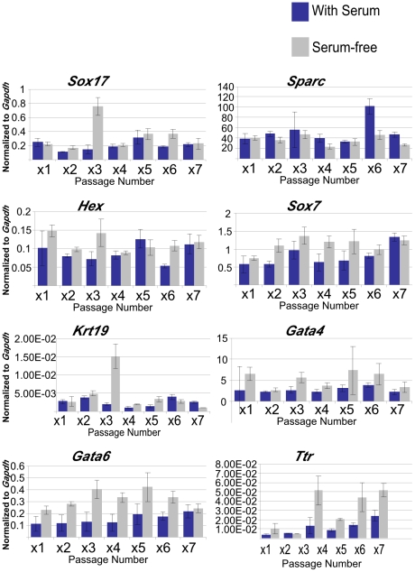
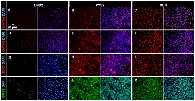
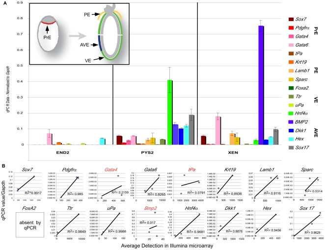
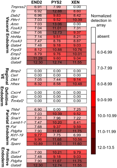
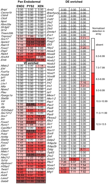
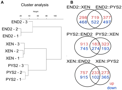
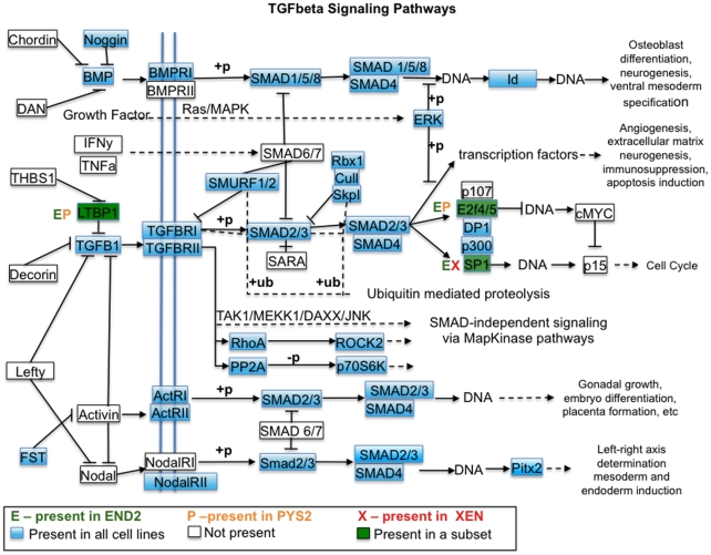
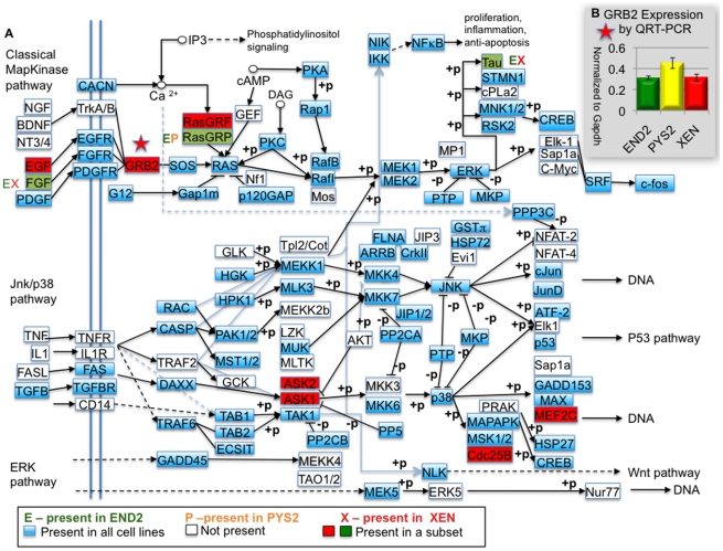
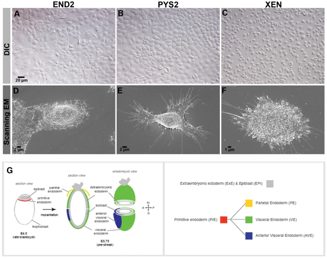
Prior to gastrulation in the mouse, all endodermal cells arise from the primitive endoderm of the blastocyst stage embryo. Primitive endoderm and its derivatives are generally referred to as extra-embryonic endoderm (ExEn) because the majority of these cells contribute to extra-embryonic lineages encompassing the visceral endoderm (VE) and the parietal endoderm (PE). During gastrulation, the definitive endoderm (DE) forms by ingression of cells from the epiblast. The DE comprises most of the cells of the gut and its accessory organs. Despite their different origins and fates, there is a surprising amount of overlap in marker expression between the ExEn and DE, making it difficult to distinguish between these cell types by marker analysis. This is significant for two main reasons. First, because endodermal organs, such as the liver and pancreas, play important physiological roles in adult animals, much experimental effort has been directed in recent years toward the establishment of protocols for the efficient derivation of endodermal cell types in vitro. Conversely, factors secreted by the VE play pivotal roles that cannot be attributed to the DE in early axis formation, heart formation and the patterning of the anterior nervous system. Thus, efforts in both of these areas have been hampered by a lack of markers that clearly distinguish between ExEn and DE. To further understand the ExEn we have undertaken a comparative analysis of three ExEn-like cell lines (END2, PYS2 and XEN). PYS2 cells are derived from embryonal carcinomas (EC) of 129 strain mice and have been characterized as parietal endoderm-like [1], END2 cells are derived from P19 ECs and described as visceral endoderm-like, while XEN cells are derived from blastocyst stage embryos and are described as primitive endoderm-like. Our analysis suggests that none of these cell lines represent a bona fide single in vivo lineage. Both PYS2 and XEN cells represent mixed populations expressing markers for several ExEn lineages. Conversely END2 cells, which were previously characterized as VE-like, fail to express many markers that are widely expressed in the VE, but instead express markers for only a subset of the VE, the anterior visceral endoderm. In addition END2 cells also express markers for the PE. We extended these observations with microarray analysis which was used to probe and refine previously published data sets of genes proposed to distinguish between DE and VE. Finally, genome-wide pathway analysis revealed that SMAD-independent TGFbeta signaling through a TAK1/p38/JNK or TAK1/NLK pathway may represent one mode of intracellular signaling shared by all three of these lines, and suggests that factors downstream of these pathways may mediate some functions of the ExEn. These studies represent the first step in the development of XEN cells as a powerful molecular genetic tool to study the endodermal signals that mediate the important developmental functions of the extra-embryonic endoderm. Our data refine our current knowledge of markers that distinguish various subtypes of endoderm. In addition, pathway analysis suggests that the ExEn may mediate some of its functions through a non-classical MAP Kinase signaling pathway downstream of TAK1.
在小鼠原肠胚形成之前,所有内胚层细胞都来源于囊胚期胚胎的原始内胚层。原始内胚层及其衍生物通常被称为胚外内胚层(ExEn),因为这些细胞的大多数都有助于包含内脏内胚层(VE)和滋养外胚层(PE)的胚外谱系。在原肠胚形成过程中,来自上胚层的细胞内陷形成原肠胚内胚层(DE)。DE 由肠道及其附属器官的大部分细胞组成。尽管它们的起源和命运不同,但 ExEn 和 DE 之间的标记物表达有惊人的重叠,这使得通过标记物分析很难区分这两种细胞类型。这有两个主要原因。首先,由于内胚层器官(如肝脏和胰腺)在成年动物中发挥着重要的生理作用,近年来,人们一直在努力建立有效的体外内胚层细胞类型的衍生方法。相反,VE 分泌的因子在早期轴形成、心脏形成和前神经系统的模式形成中起着关键作用,这些作用不能归因于 DE。因此,这两个领域的努力都因缺乏能明确区分 ExEn 和 DE 的标记物而受到阻碍。为了进一步了解 ExEn,我们对三种 ExEn 样细胞系(END2、PYS2 和 XEN)进行了比较分析。PYS2 细胞来源于 129 品系小鼠的胚胎癌细胞(EC),并被特征化为滋养外胚层样[1],END2 细胞来源于 P19 ECs,被描述为内脏内胚层样,而 XEN 细胞来源于囊胚期胚胎,被描述为原始内胚层样。我们的分析表明,这些细胞系都没有一个真正的单一体内谱系。PYS2 和 XEN 细胞都代表混合群体,表达几种 ExEn 谱系的标记物。相反,以前被描述为 VE 样的 END2 细胞未能表达广泛存在于 VE 中的许多标记物,而只表达 VE 的一小部分标记物,即前内脏内胚层。此外,END2 细胞还表达滋养外胚层的标记物。我们通过微阵列分析扩展了这些观察结果,该分析用于探测和完善先前发表的用于区分 DE 和 VE 的基因数据集。最后,全基因组途径分析表明,SMAD 非依赖性 TGFβ信号通过 TAK1/p38/JNK 或 TAK1/NLK 途径可能代表这三种途径共有的一种细胞内信号传导模式,并表明这些途径下游的因子可能介导 ExEn 的一些功能。这些研究代表了将 XEN 细胞作为研究介导胚外内胚层重要发育功能的内胚层信号的强大分子遗传工具的第一步。我们的数据细化了我们当前对区分各种内胚层亚型的标记物的认识。此外,途径分析表明,ExEn 可能通过 TAK1 下游的非经典 MAP 激酶信号通路来介导其部分功能。