Department of Cancer Biology, Vanderbilt University School of Medicine , Nashville, TN 37232 , USA.
Biol Open. 2012 Aug 15;1(8):711-22. doi: 10.1242/bio.20121867. Epub 2012 Jun 12.
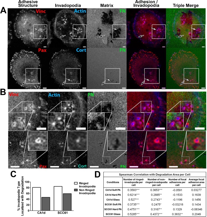
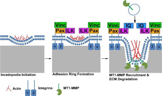
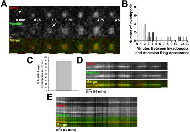
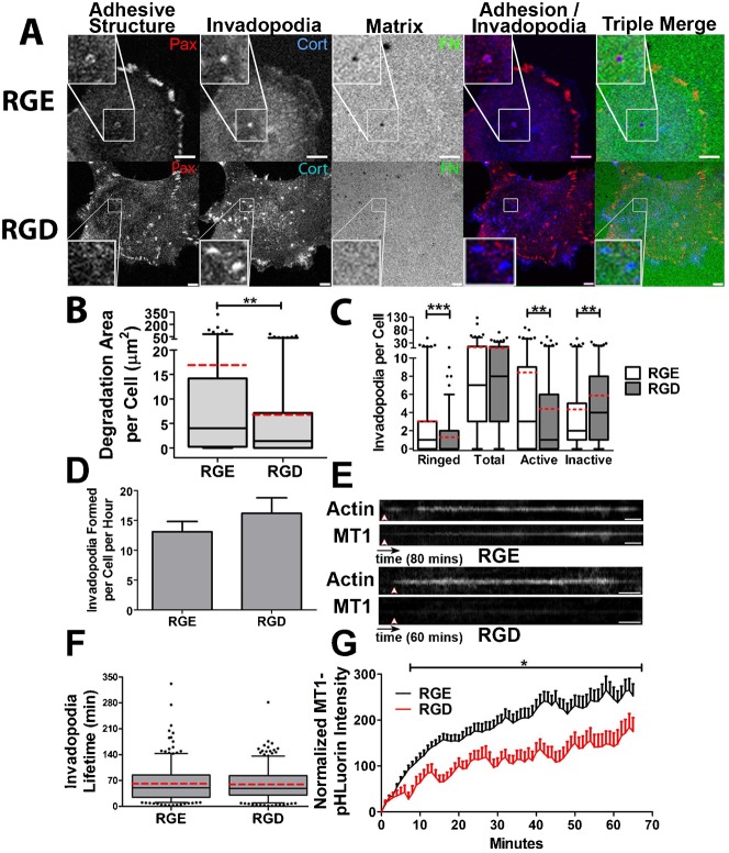
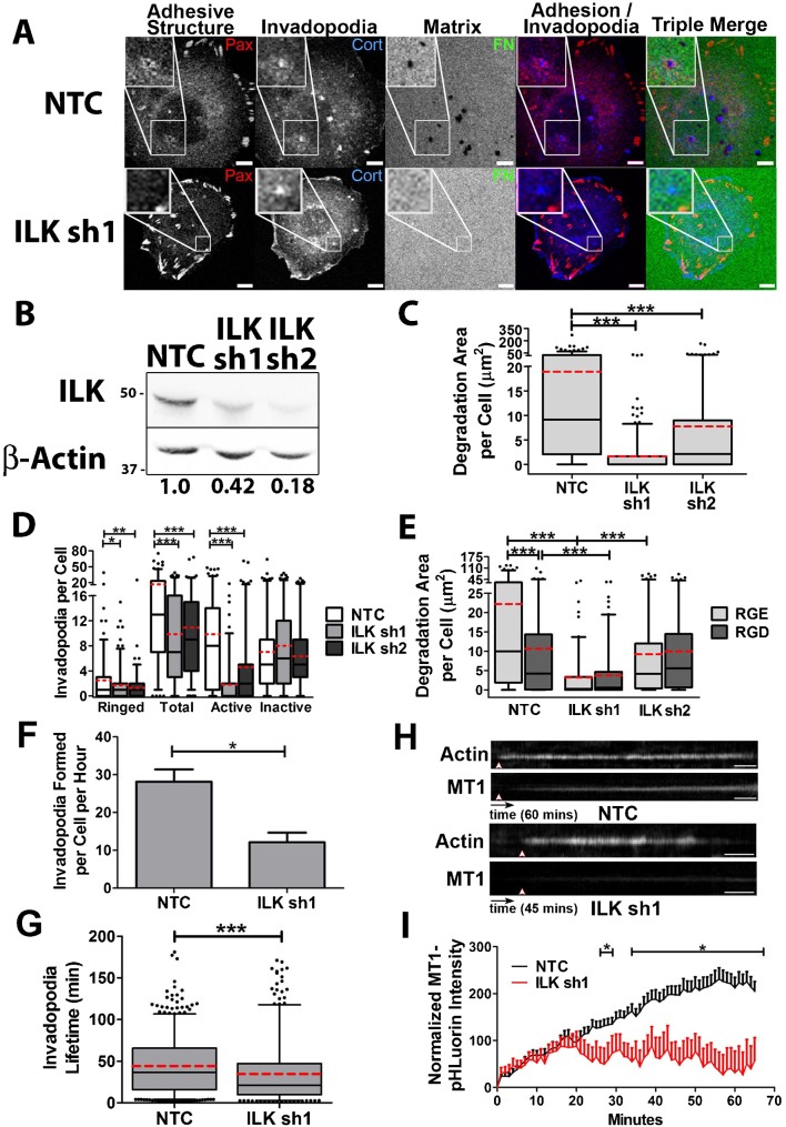
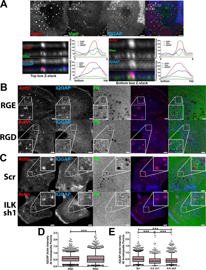
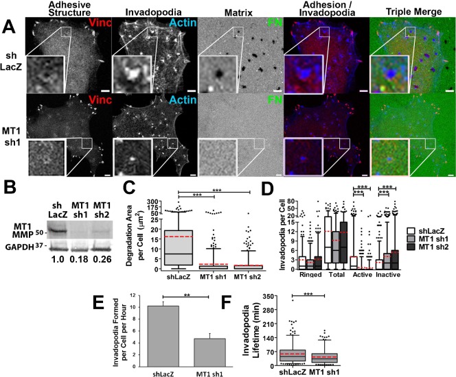
Invasion and metastasis are aggressive cancer phenotypes that are highly related to the ability of cancer cells to degrade extracellular matrix (ECM). At the cellular level, specialized actin-rich structures called invadopodia mediate focal matrix degradation by serving as exocytic sites for ECM-degrading proteinases. Adhesion signaling is likely to be a critical regulatory input to invadopodia, but the mechanism and location of such adhesion signaling events are poorly understood. Here, we report that adhesion rings surround invadopodia shortly after formation and correlate strongly with invadopodium activity on a cell-by-cell basis. By contrast, there was little correlation of focal adhesion number or size with cellular invadopodium activity. Prevention of adhesion ring formation by inhibition of RGD-binding integrins or knockdown (KD) of integrin-linked kinase (ILK) reduced the number of ECM-degrading invadopodia and reduced recruitment of IQGAP to invadopodium actin puncta. Furthermore, live cell imaging revealed that the rate of extracellular MT1-MMP accumulation at invadopodia was greatly reduced in both integrin-inhibited and ILK-KD cells. Conversely, KD of MT1-MMP reduced invadopodium activity and dynamics but not the number of adhesion-ringed invadopodia. These results suggest a model in which adhesion rings are recruited to invadopodia shortly after formation and promote invadopodium maturation by enhancing proteinase secretion. Since adhesion rings are a defining characteristic of podosomes, similar structures formed by normal cells, our data also suggest further similarities between invadopodia and podosomes.
侵袭和转移是具有侵略性的癌症表型,与癌细胞降解细胞外基质(ECM)的能力高度相关。在细胞水平上,称为侵袭伪足的特化的富含肌动蛋白的结构通过充当 ECM 降解蛋白酶的胞吐位点来介导局灶性基质降解。黏附信号可能是侵袭伪足的关键调节输入,但这种黏附信号事件的机制和位置知之甚少。在这里,我们报告说,黏附环在侵袭伪足形成后不久就围绕着侵袭伪足,并与侵袭伪足在细胞基础上的活性密切相关。相比之下,黏附斑的数量或大小与细胞侵袭伪足活性几乎没有相关性。通过抑制 RGD 结合整联蛋白或整联蛋白连接激酶(ILK)的 KD 来阻止黏附环的形成,减少了 ECM 降解侵袭伪足的数量,并减少了 IQGAP 向侵袭伪足肌动蛋白点状结构的募集。此外,活细胞成像显示,在整联蛋白抑制和 ILK-KD 细胞中,细胞外 MT1-MMP 的积累速度在侵袭伪足处大大降低。相反,MT1-MMP 的 KD 降低了侵袭伪足的活性和动力学,但没有减少黏附环侵袭伪足的数量。这些结果表明,黏附环在侵袭伪足形成后不久就被募集到侵袭伪足,并通过增强蛋白酶分泌来促进侵袭伪足的成熟。由于黏附环是足状伪足的一个特征,正常细胞也形成类似的结构,因此我们的数据还表明侵袭伪足和足状伪足之间存在进一步的相似性。