Department of Gastroenterology and Hepatology, Okayama University Graduate School of Medicine, Dentistry, and Pharmaceutical Sciences, Okayama, Japan.
Biomed Eng Online. 2012 Dec 7;11:93. doi: 10.1186/1475-925X-11-93.
Bioartificial liver systems, designed to support patients with liver failure, are composed of bioreactors and functional hepatocytes. Immunological rejection of the embedded hepatocytes by the host immune system is a serious concern that crucially degrades the performance of the device. Induced pluripotent stem (iPS) cells are considered a desirable source for bioartificial liver systems, because patient-derived iPS cells are free from immunological rejection. The purpose of this paper was to test the feasibility of a bioartificial liver system with iPS cell-derived hepatocyte-like cells.
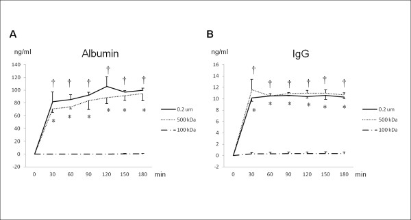
Mouse iPS cells were differentiated into hepatocyte-like cells by a multi-step differentiation protocol via embryoid bodies and definitive endoderm. Differentiation of iPS cells was evaluated by morphology, PCR assay, and functional assays. iPS cell-derived hepatocyte-like cells were cultured in a bioreactor module with a pore size of 0.2 μm for 7 days. The amount of albumin secreted into the circulating medium was analyzed by ELISA. Additionally, after a 7-day culture in a bioreactor module, cells were observed by a scanning electron microscope.
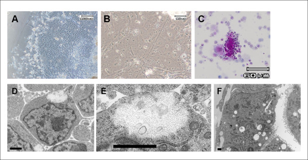
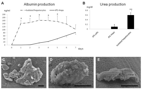
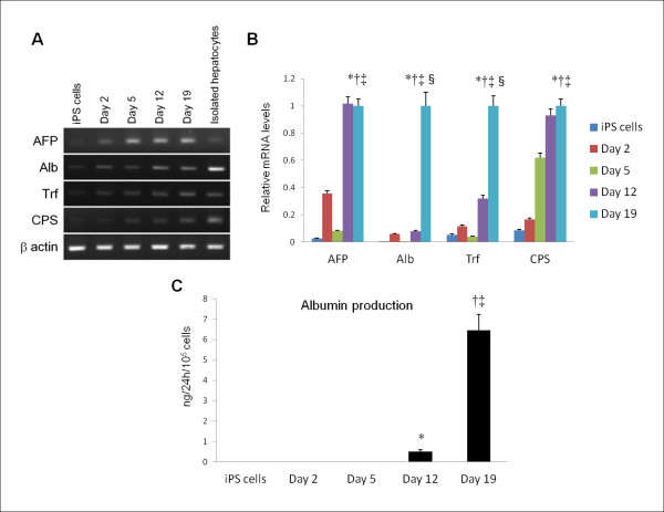
At the final stage of the differentiation program, iPS cells changed their morphology to a polygonal shape with two nucleoli and enriched cytoplasmic granules. Transmission electron microscope analysis revealed their polygonal shape, glycogen deposition in the cytoplasm, microvilli on their surfaces, and a duct-like arrangement. PCR analysis showed increased expression of albumin mRNA over the course of the differentiation program. Albumin and urea production was also observed. iPS-Heps culture in bioreactor modules showed the accumulation of albumin in the medium for up to 7 days. Scanning electron microscopy revealed the attachment of cell clusters to the hollow fibers of the module. These results indicated that iPS cells were differentiated into hepatocyte-like cells after culture for 7 days in a bioreactor module with a pore size of 0.2 μm.
We consider the combination of a bioreactor module with a 0.2-μm pore membrane and embedded hepatocytes differentiated from iPS cells to be a promising option for bioartificial liver systems. This paper provides the basic concept and preliminary data for an iPS cell-oriented bioartificial liver system.PACS code: 87. Biological and medical physics, 87.85.-d Biomedical engineering, 87.85.Lf Tissue engineering, 87.85.Tu Modeling biomedical systems.
生物人工肝脏系统旨在为肝功能衰竭的患者提供支持,它由生物反应器和功能肝细胞组成。宿主免疫系统对嵌入的肝细胞的免疫排斥是一个严重的问题,这会极大地降低设备的性能。诱导多能干细胞(iPS 细胞)被认为是生物人工肝脏系统的理想来源,因为患者来源的 iPS 细胞不会被免疫排斥。本文旨在测试基于 iPS 细胞来源的肝细胞样细胞的生物人工肝脏系统的可行性。
通过胚胎体和确定的内胚层的多步分化方案,将小鼠 iPS 细胞分化为肝细胞样细胞。通过形态学、PCR 检测和功能检测评估 iPS 细胞的分化。将 iPS 细胞衍生的肝细胞样细胞在孔径为 0.2μm 的生物反应器模块中培养 7 天。通过 ELISA 分析分析分泌到循环培养基中的白蛋白量。此外,在生物反应器模块中培养 7 天后,通过扫描电子显微镜观察细胞。
在分化方案的最后阶段,iPS 细胞的形态发生变化,呈多边形,有两个核仁,细胞质颗粒丰富。透射电子显微镜分析显示其呈多边形,细胞质中有糖原沉积,表面有微绒毛,呈导管样排列。PCR 分析显示,在分化过程中白蛋白 mRNA 的表达逐渐增加。还观察到白蛋白和尿素的产生。iPS-Heps 在生物反应器模块中的培养物在 7 天内积累白蛋白在培养基中。扫描电子显微镜显示细胞簇附着在模块的中空纤维上。这些结果表明,iPS 细胞在孔径为 0.2μm 的生物反应器模块中培养 7 天后可分化为肝细胞样细胞。
我们认为,生物反应器模块与 0.2μm 孔径的膜以及从 iPS 细胞分化而来的嵌入式肝细胞的结合,是生物人工肝脏系统的一种很有前途的选择。本文为基于 iPS 细胞的生物人工肝脏系统提供了基本概念和初步数据。
PACS 码:87. 生物和医学物理学,87.85.d 生物医学工程,87.85.Lf 组织工程学,87.85.Tu 生物医学系统建模。