Szopiński Tomasz R, Sudoł-Szopińska Iwona, Furmanek Mariusz I, Dzik Tomasz, Chłosta Piotr L, Borówka Andrzej
Department of Urology, Postgraduate Medical Educational Centre, Warsaw, Poland.
Wideochir Inne Tech Maloinwazyjne. 2012 Mar;7(1):55-8. doi: 10.5114/wiitm.2011.25622. Epub 2011 Nov 8.
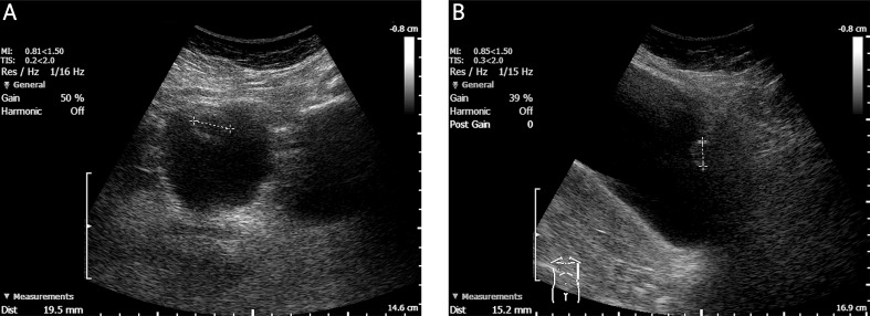
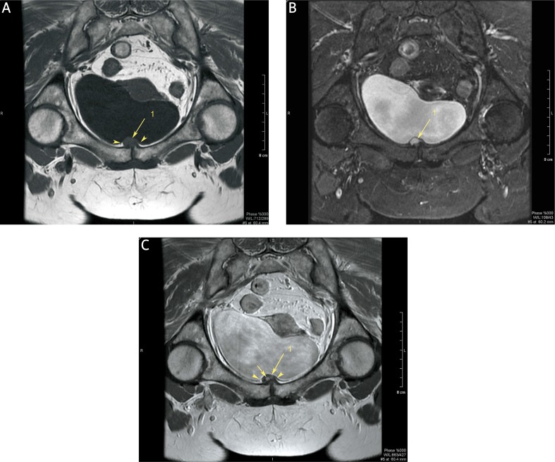
Urinary bladder sonography is a sensitive diagnostic technique used for visualizing urinary bladder tumours. The aim of our communication is to present a case of a pseudotumour of the urinary bladder originating from the symphysis pubis syndesmosis. A 58-year-old woman was seen by a urologist with symptoms of lower urinary tract infection. Urinary bladder sonography was performed, followed by magnetic resonance imaging. Sonographic images of the bladder showed an exophytic mass on the urinary bladder's anterior wall. A transurethral resection of the tumour was performed. A histopathological examination revealed a necrotic extramural mass, without traits of malignancy. The mass reappeared in the follow-up vesical sonography. Subsequently, its transurethral resection was repeated with the same histopathological findings. The next urinary bladder sonography revealed the presence of the mass again. Pelvic magnetic resonance imaging was performed, which showed advanced degenerative changes in the pubic symphysis syndesmosis that protruded into the bladder, imitating a urinary bladder tumour. To avoid unnecessary surgery, both radiologists and urologists should be made aware that there is a possibility of similar cases in patients. Magnetic resonance imaging enabled correct determination of the primary site of the growth, which, together with the histopathological examination results, influenced the choice of the implemented therapeutic procedures.
膀胱超声检查是一种用于可视化膀胱肿瘤的敏感诊断技术。我们此次交流的目的是呈现一例起源于耻骨联合韧带的膀胱假瘤病例。一名58岁女性因下尿路感染症状就诊于泌尿科医生。进行了膀胱超声检查,随后进行了磁共振成像。膀胱超声图像显示膀胱前壁有一个外生性肿块。对该肿瘤进行了经尿道切除术。组织病理学检查显示为坏死性壁外肿块,无恶性特征。在后续的膀胱超声检查中肿块再次出现。随后,再次进行经尿道切除术,组织病理学结果相同。下一次膀胱超声检查再次显示肿块存在。进行了盆腔磁共振成像,结果显示耻骨联合韧带出现严重退行性改变并突入膀胱,形似膀胱肿瘤。为避免不必要的手术,放射科医生和泌尿科医生均应意识到患者有可能出现类似病例。磁共振成像能够正确确定病变的原发部位,这与组织病理学检查结果共同影响了所实施治疗方案的选择。