Medical Research Center of Shandong Provincial Qianfoshan Hospital, Shandong University, Jingshi road 16766, Jinan, Shandong 250014, PR China.
BMC Musculoskelet Disord. 2012 Dec 20;13:256. doi: 10.1186/1471-2474-13-256.
Studies have demonstrated that carbonic anhydrase I (CA1) stimulates calcium salt precipitation and cell calcification, which is an essential step in new bone formation. Our study had reported that CA1 encoding gene has a strong association with rheumatoid arthritis (RA) and ankylosing spondylitis (AS), two rheumatic diseases with abnormal new bone formation and bone resorption in joints. This study investigated the effect of CA1 on joint inflammation and tissue destruction in transgenic mice that over-express CA1 (CA1-Tg).
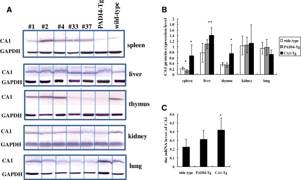
CA1-Tg was generated with C57BL/6J mice by conventional methods. CA1-Tg was treated with collagen-II to induce arthritis (CIA). Wild-type mice, CA1-Tg treated with bovine serum albumin (BSA) and transgenic mice over-expressing PADI4 (PADI4-Tg), a gene known to be involved in rheumatoid arthritis, were used as controls. Histochemistry and X-ray radiographic assay were used to examine joint destruction. Western blotting and real time-PCR were used to examine CA1 expression.
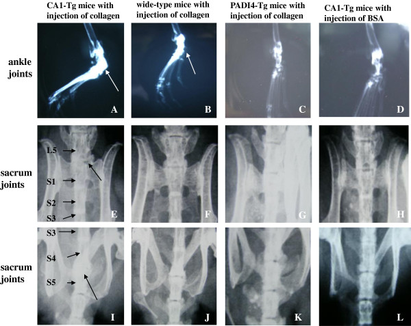
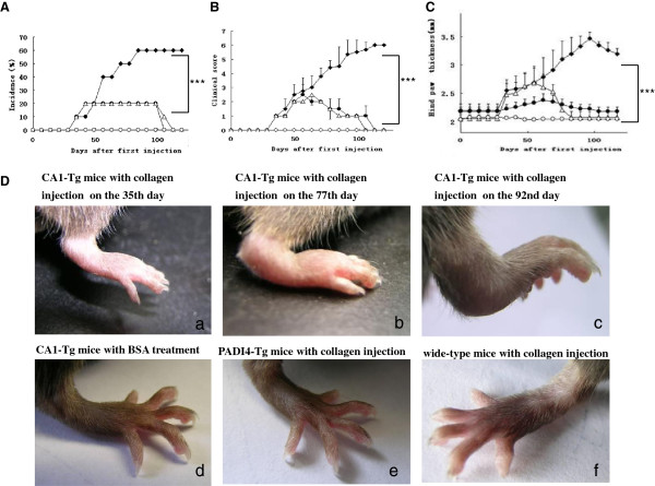
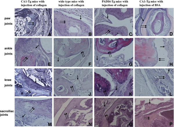
CIA was observed in 60% of CA1-Tg, 20% of PADI4-Tg and 20% of wild-type mice after collagen injections. No CIA was found in CA1-Tg mice that received injections of BSA. The arthritic score was 5.5 ± 0.84 in the CA1-Tgs but the score was less than 2 in the injected wild-type mice and the PADI4-Tgs. The thickness of the hind paws in the CA1-Tgs was 3.46 ± 0.11 mm, which was thicker than that of PADI4-Tgs (2.23 ± 0.08 mm), wild-type mice (2.08 ± 0.06 mm) and BSA-treated CA1-Tgs (2.04 ± 0.07 mm). Histochemistry showed obvious inflammation, synovial hyperplasia and bone destruction in the joints of CA1-Tg that was not detected in PADI4-Tgs or wild-type mice. X-ray assays showed bone fusion in the paws and spines of CA1-Tg mice.
Over-expression of CA1 may aggravate joint inflammation and tissue destruction in the transgenic mice.
研究表明碳酸酐酶 I(CA1)可刺激钙盐沉淀和细胞钙化,这是新骨形成的关键步骤。我们的研究报告称,CA1 编码基因与类风湿关节炎(RA)和强直性脊柱炎(AS)密切相关,这两种风湿性疾病在关节中均存在异常的新骨形成和骨吸收。本研究探讨了过表达 CA1(CA1-Tg)的转基因小鼠中 CA1 对关节炎症和组织破坏的影响。
通过常规方法用 C57BL/6J 小鼠生成 CA1-Tg。用胶原蛋白 II 处理 CA1-Tg 以诱导关节炎(CIA)。将野生型小鼠、用牛血清白蛋白(BSA)处理的 CA1-Tg 和过表达已知与类风湿关节炎相关的基因 PADI4(PADI4-Tg)的转基因小鼠作为对照。采用组织化学和 X 射线放射照相检测关节破坏。采用 Western blot 和实时 PCR 检测 CA1 表达。
在胶原注射后,60%的 CA1-Tg、20%的 PADI4-Tg 和 20%的野生型小鼠出现 CIA。接受 BSA 注射的 CA1-Tg 未出现 CIA。CA1-Tg 的关节炎评分为 5.5±0.84,而注射的野生型小鼠和 PADI4-Tg 的评分小于 2。CA1-Tg 的后爪厚度为 3.46±0.11mm,明显大于 PADI4-Tg(2.23±0.08mm)、野生型小鼠(2.08±0.06mm)和 BSA 处理的 CA1-Tg(2.04±0.07mm)。组织化学显示 CA1-Tg 关节中有明显的炎症、滑膜增生和骨破坏,但在 PADI4-Tg 或野生型小鼠中未检测到。X 射线检测显示 CA1-Tg 小鼠的爪子和脊柱有骨融合。
CA1 的过表达可能会加重转基因小鼠的关节炎症和组织破坏。