Tur Carmen, Wheeler-Kingshott Claudia A M, Altmann Daniel R, Miller David H, Thompson Alan J, Ciccarelli Olga
Department of Brain Repair and Rehabilitation, UCL Institute of Neurology, London, United Kingdom; Department of Medicine, Clinical Neuroimmunology Unit, Multiple Sclerosis Centre of Catalonia (CEM-Cat), Autonomous University of Barcelona, CARM-Vall d'Hebron University Hospital, Barcelona, Spain.
Hum Brain Mapp. 2014 Mar;35(3):993-1003. doi: 10.1002/hbm.22229. Epub 2012 Dec 26.
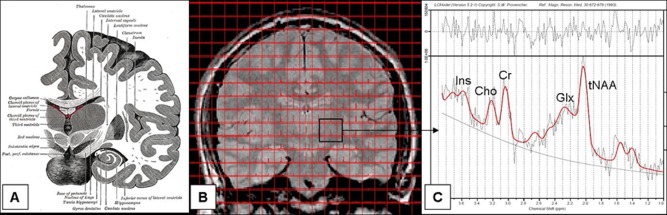
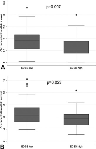
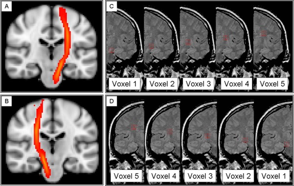
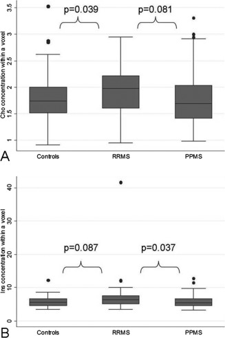
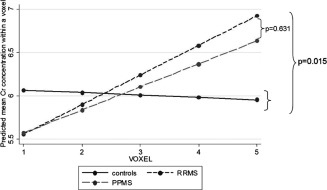
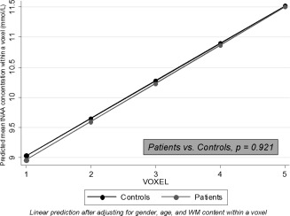
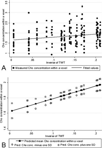
We characterized metabolic changes along the cortico-spinal tract (CST) in multiple sclerosis (MS) patients using a novel application of chemical shift imaging (CSI) and considering the spatial variation of metabolite levels. Thirteen relapsing-remitting (RR) and 13 primary-progressive (PP) MS patients and 16 controls underwent (1)H-MR CSI, which was applied to coronal-oblique scans to sample the entire CST. The concentrations of the main metabolites, i.e., N-acetyl-aspartate, myo-Inositol (Ins), choline containing compounds (Cho) and creatine and phosphocreatine (Cr), were calculated within voxels placed in regions where the CST is located, from cerebral peduncle to corona radiata. Differences in metabolite concentrations between groups and associations between metabolite concentrations and disability were investigated, allowing for the spatial variability of metabolite concentrations in the statistical model. RRMS patients showed higher CST Cho concentration than controls, and higher CST Ins concentration than PPMS, suggesting greater inflammation and glial proliferation in the RR than in the PP course. In RRMS, a significant, albeit modest, association between greater Ins concentration and greater disability suggested that gliosis may be relevant to disability. In PPMS, lower CST Cho and Cr concentrations correlated with greater disability, suggesting that in the progressive stage of the disease, inflammation declines and energy metabolism reduces. Attention to the spatial variation of metabolite concentrations made it possible to detect in patients a greater increase in Cr concentration towards the superior voxels as compared to controls and a stronger association between Cho and disability, suggesting that this step improves our ability to identify clinically relevant metabolic changes.
我们使用化学位移成像(CSI)的一种新应用,并考虑代谢物水平的空间变化,对多发性硬化症(MS)患者沿皮质脊髓束(CST)的代谢变化进行了表征。13名复发缓解型(RR)和13名原发进展型(PP)MS患者以及16名对照者接受了氢质子磁共振化学位移成像(¹H-MR CSI),该成像应用于冠状斜位扫描以对整个CST进行采样。在位于从大脑脚到放射冠的CST所在区域的体素内,计算主要代谢物的浓度,即N-乙酰天门冬氨酸、肌醇(Ins)、含胆碱化合物(Cho)以及肌酸和磷酸肌酸(Cr)。研究了组间代谢物浓度的差异以及代谢物浓度与残疾之间的关联,并在统计模型中考虑了代谢物浓度的空间变异性。RRMS患者的CST中Cho浓度高于对照组,CST中Ins浓度高于PPMS患者,这表明RR病程中的炎症和胶质细胞增殖比PP病程更严重。在RRMS中,Ins浓度升高与残疾程度增加之间存在显著(尽管适度)关联,这表明胶质增生可能与残疾有关。在PPMS中,CST中较低的Cho和Cr浓度与更严重的残疾相关,这表明在疾病的进展阶段,炎症减轻且能量代谢降低。关注代谢物浓度的空间变化使得能够检测到患者中与对照组相比,Cr浓度朝着上方体素的增加更大,以及Cho与残疾之间的关联更强,这表明这一步骤提高了我们识别临床相关代谢变化的能力。