Division of Cardiovascular Medicine, Department of Internal Medicine, Kobe University Graduate School of Medicine, 7-5-1 Kusunoki-cho, Chuo-ku, Kobe, Hyogo 650-0017, Japan.
Eur Heart J Cardiovasc Imaging. 2013 Sep;14(9):865-75. doi: 10.1093/ehjci/jes299. Epub 2013 Jan 4.
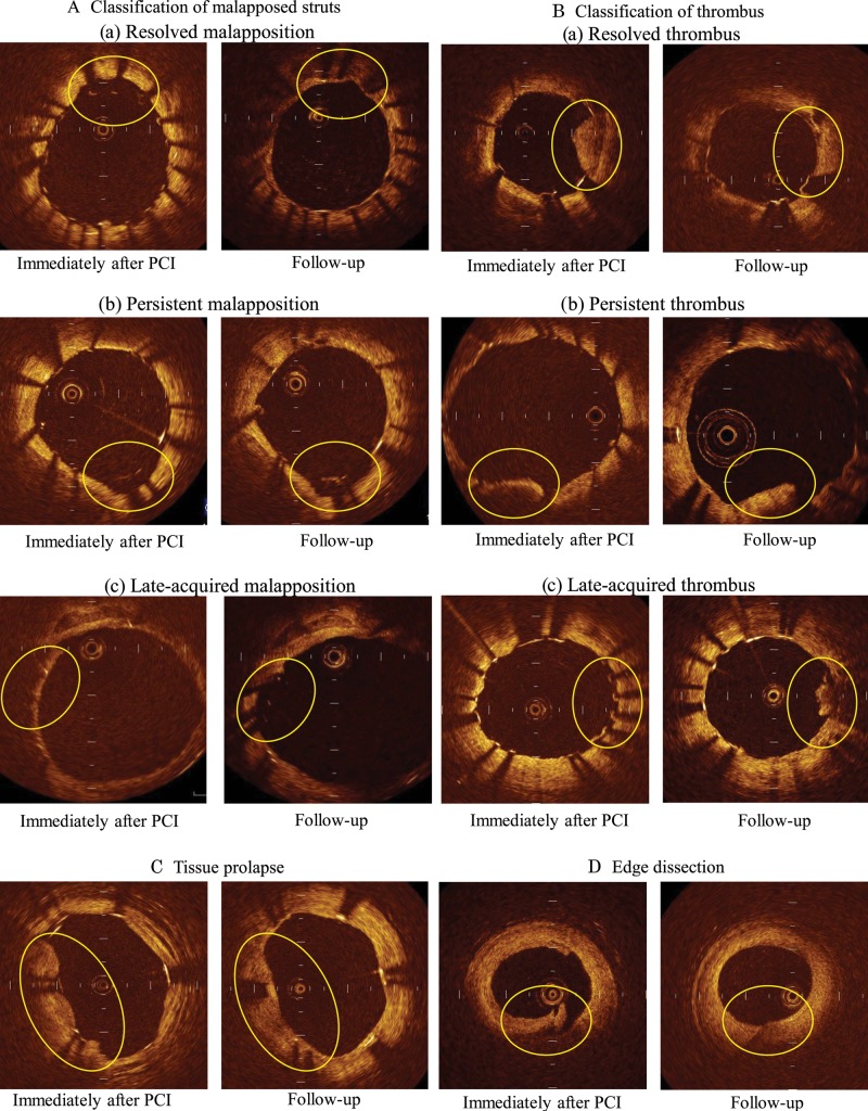
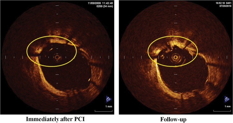
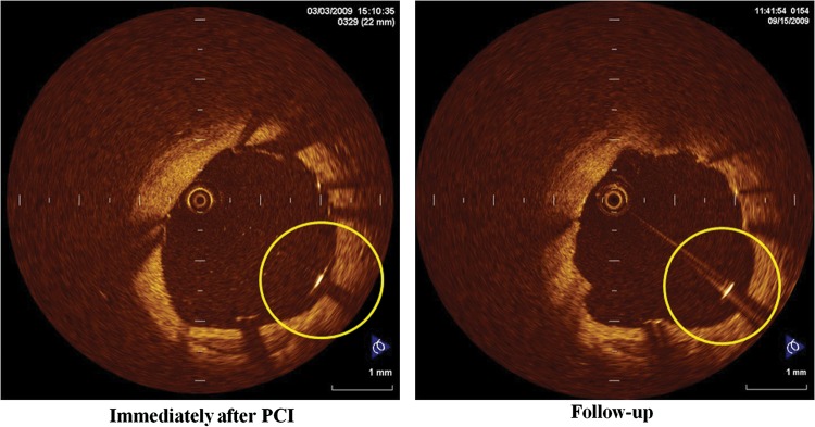
We performed this study to clarify natural consequences of abnormal structures (stent malapposition, thrombus, tissue prolapse, and stent edge dissection) after percutaneous coronary intervention (PCI).
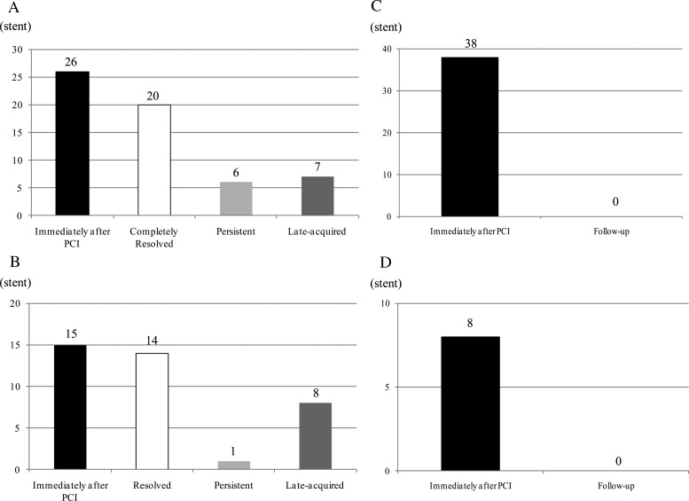
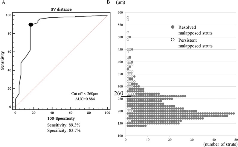
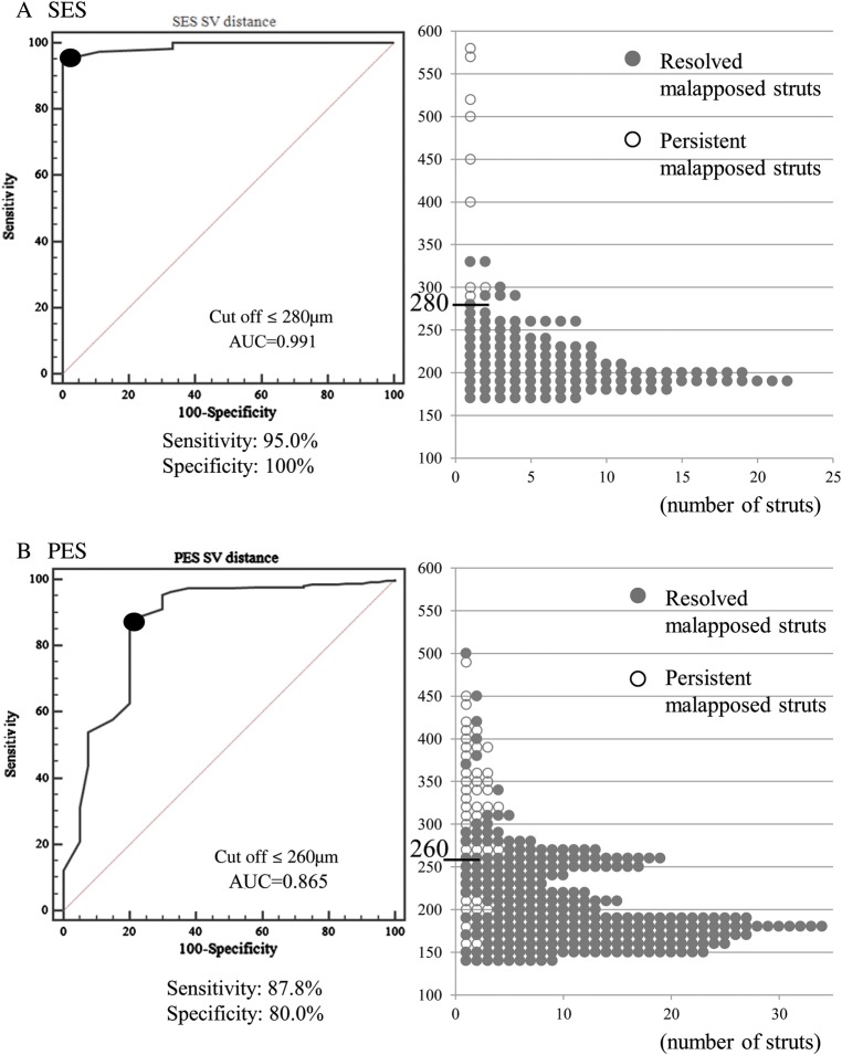
Thirty-five patients treated with 40 drug-eluting stents underwent serial optical coherence tomography (OCT) imaging immediately after PCI and at the 8-month follow-up. Among a total of 73 929 struts in every frame, 431 struts (26 stents) showed malapposition immediately after PCI. Among these, 49 remained malapposed at the follow-up examination. The mean distance between the strut and vessel wall (S-V distance) of persistent malapposed struts on post-stenting OCT images was significantly longer than that of resolved malapposed struts (342 ± 99 vs. 210 ± 49 μm; P <0.01). Based on receiver-operating characteristic curve analysis, an S-V distance ≤260 µm on post-stenting OCT images was the corresponding cut-off point for resolved malapposed struts (sensitivity: 89.3%, specificity: 83.7%, area under the curve = 0.884). Additionally, 108 newly appearing malapposed struts were observed on follow-up OCT, probably due to thrombus dissolution or plaque regression. Thrombus was observed in 15 stents post-PCI. Serial OCT analysis revealed persistent thrombus in 1 stent, resolved thrombus in 14 stents, and late-acquired thrombus in 8 stents. Tissue prolapse observed in 38 stents had disappeared at the follow-up. All eight stent edge dissections were repaired at the follow-up.
Most cases of stent malapposition with a short S-V distance, thrombus, tissue prolapse, or minor stent edge dissection improved during the follow-up. These OCT-detected minor abnormalities may not require additional treatment.
本研究旨在阐明经皮冠状动脉介入治疗(PCI)后异常结构(支架贴壁不良、血栓、组织脱垂和支架边缘夹层)的自然转归。
35 例接受 40 枚药物洗脱支架治疗的患者在 PCI 后即刻和 8 个月随访时接受了连续光学相干断层扫描(OCT)成像。在每一帧的总共 73929 个支架中,431 个支架(26 个支架)在 PCI 后即刻出现贴壁不良。其中,49 个在随访检查时仍存在贴壁不良。支架置入后 OCT 图像上持续贴壁不良的支架的支架-血管壁距离(S-V 距离)明显长于已解决的贴壁不良的支架(342±99 μm比 210±49 μm;P<0.01)。基于受试者工作特征曲线分析,支架置入后 OCT 图像上的 S-V 距离≤260 μm 是解决贴壁不良的相应截断点(敏感性:89.3%,特异性:83.7%,曲线下面积=0.884)。此外,在随访 OCT 上观察到 108 个新出现的贴壁不良的支架,可能是由于血栓溶解或斑块消退。支架置入后发现 15 个支架内血栓形成。连续 OCT 分析显示,1 个支架内持续存在血栓,14 个支架内血栓溶解,8 个支架内迟发性血栓形成。在 38 个支架中观察到的组织脱垂在随访时已消失。所有 8 个支架边缘夹层均在随访时修复。
支架贴壁不良、血栓形成、组织脱垂或轻微支架边缘夹层,伴有短 S-V 距离的大多数病例在随访期间得到改善。这些 OCT 检测到的轻微异常可能不需要额外治疗。